Your cart
There are no more items in your cart

Created using ultra-high-resolution 3D color printing.
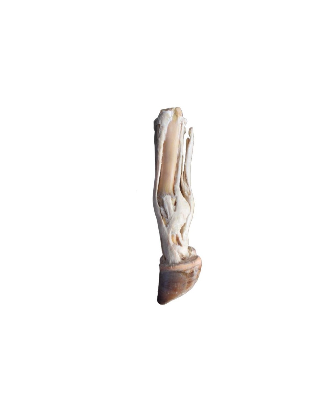
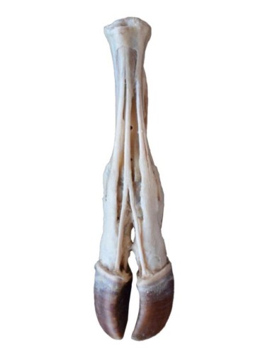
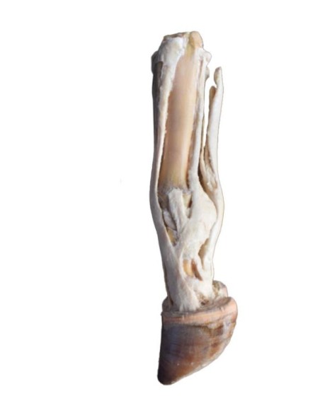
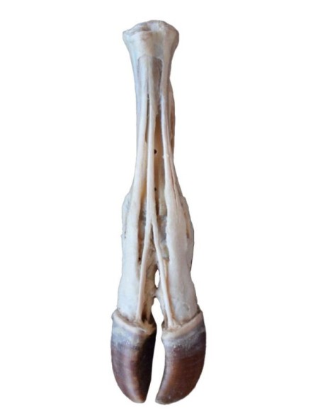
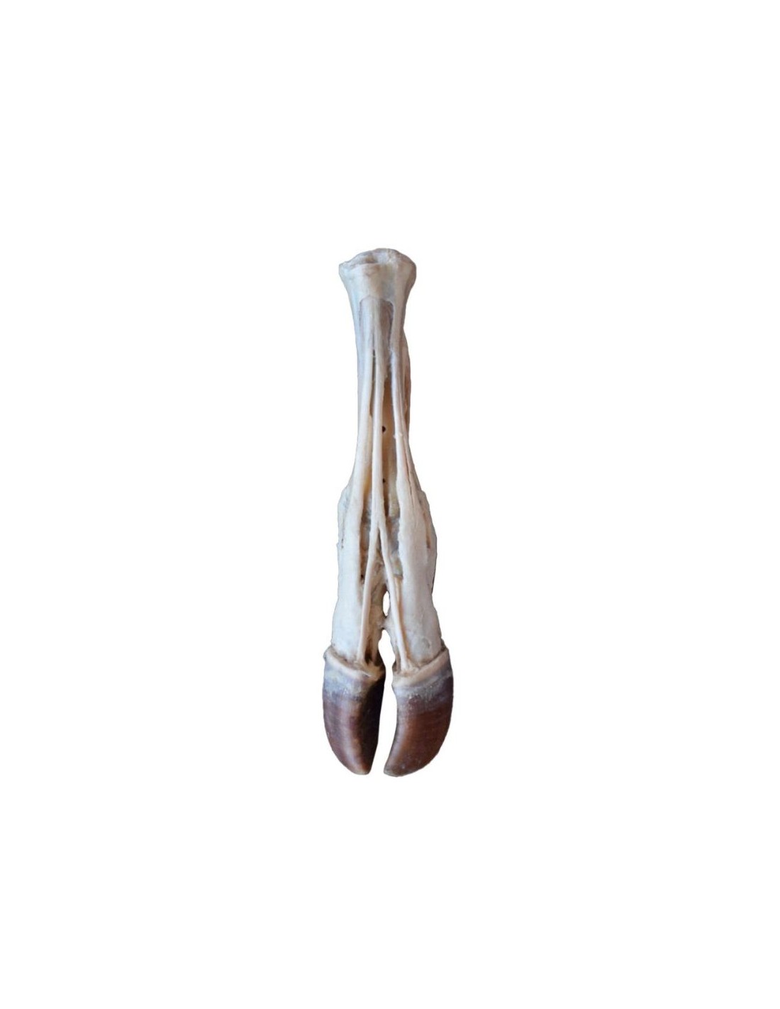
Ox Foot – Tendons and Ligaments VP11000
This model is part of the exclusive Monash 3D Anatomy series, a comprehensive collection of human dissections reproduced using high-resolution color 3D printing.
Product Description
The Ox Foot – Tendons and Ligaments (VP11000) anatomical model is designed to provide a detailed and realistic view of the tendon and ligament structures of the bovine foot. Thanks to this accurate representation, veterinary students and professionals can deepen their understanding of the biomechanics and anatomy of the ox foot. Ideal for anatomy courses, dissection labs, and research activities, the VP11000 is an essential teaching tool with significant educational impact.
Key Features
Detailed and Realistic Anatomy
- Faithful Reproduction: The model highlights the tendons and ligaments of the bovine foot, allowing for an in-depth study of the structures involved in support and movement.
- Educational Focus: Every element is carefully designed to facilitate understanding of fundamental anatomical relationships and the connections between different tissues.
High-Quality Materials
- Robust Construction: Made with premium materials, the VP11000 ensures strength and durability, making it suitable for intensive use in educational and research settings.
- Precision Finishes: Every detail has been carefully crafted to offer the most realistic educational experience possible.
Wide Range of Educational and Research Applications
- Advanced Educational Tool: Ideal for veterinary anatomy courses, seminars, and workshops, the model allows students to acquire practical skills and reinforce theoretical knowledge.
- Research Support: Perfect for comparative studies and analyses of the bovine foot, helping to enhance the skills of those working in the livestock and veterinary fields.
Benefits of Use
Interactive and Safe Learning
With the Ox Hoof – Tendons and Ligaments model (VP11000), students and professionals can practice ethically and safely, eliminating the need to use live animals. This tool allows for a thorough understanding of the biomechanics and structure of the bovine hoof.
Investment for Institutions and Training Centers
This model represents a strategic opportunity for universities, veterinary schools, and research centers, enhancing the effectiveness of teaching and practical training through an accurate anatomical representation.
Technical Specifications
For further information regarding dimensions, materials used, and instructions for use, please consult the complete technical data sheet or contact our customer service for personalized support.
Buy Now
Don’t miss the chance to enhance your education or that of your students with the Ox Foot – Tendons and Ligaments
model (VP11000)
.
Add to cart
and discover how this tool can revolutionize the understanding and study of bovine anatomy.
What advantages does the Monash University anatomical dissection collection offer compared to plastic models or plastinated specimens?
- Each body replica has been carefully created from selected patient radiographic data or human cadaver specimens selected by a highly qualified team of anatomists at the Monash University Centre for Human Anatomy Education to illustrate a range of clinically important areas of anatomy with a quality and fidelity that cannot be achieved with conventional anatomical models – this is real anatomy, not stylized.
- Each body replica has been rigorously checked by a team of highly qualified anatomists at the Center for Human Anatomy Education, Monash University, to ensure the anatomical accuracy of the final product.
- The body replicas are not real human tissue and are therefore not subject to any restrictions on transport, import, or use in educational institutions that do not hold an anatomy license. The Monash 3D Anatomy dissection series avoids these and other ethical issues that arise when dealing with plastinated human remains.
No reviews
Tap to zoom