Your cart
There are no more items in your cart


Deep Palmar Dissection of the Horse's Foot VP10040
erler zimmer
EZ-VP10040
Out-of-Stock
€1,990.00
Tax included
Created using ultra-high-resolution 3D color printing.
Deep Palmar Dissection of the Horse's Foot VP10040
This model is part of the exclusive Monash 3D Anatomy series, a comprehensive collection of human dissections reproduced using high-resolution color 3D printing.
Product Description
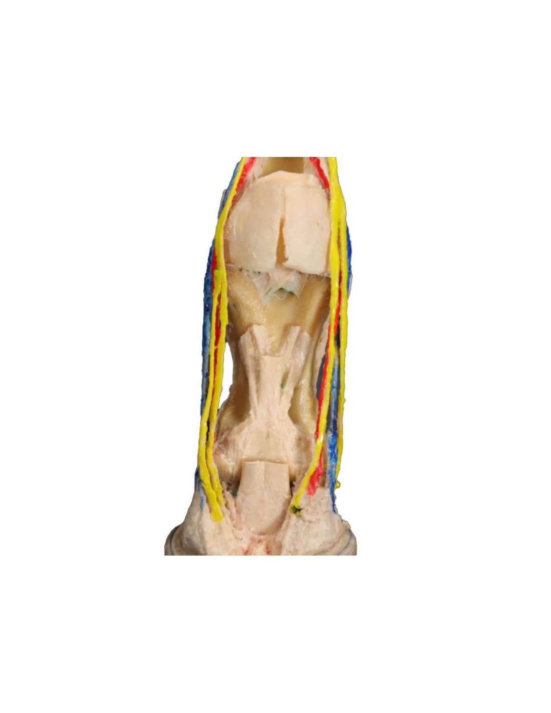
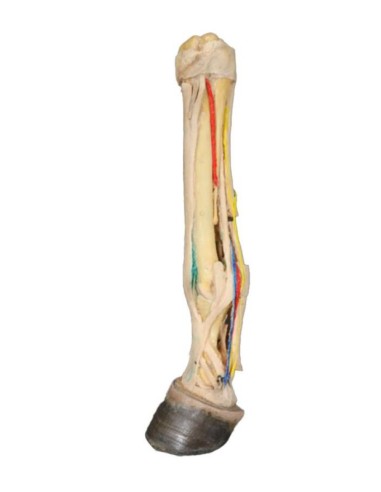
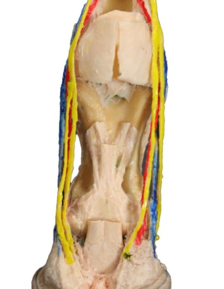
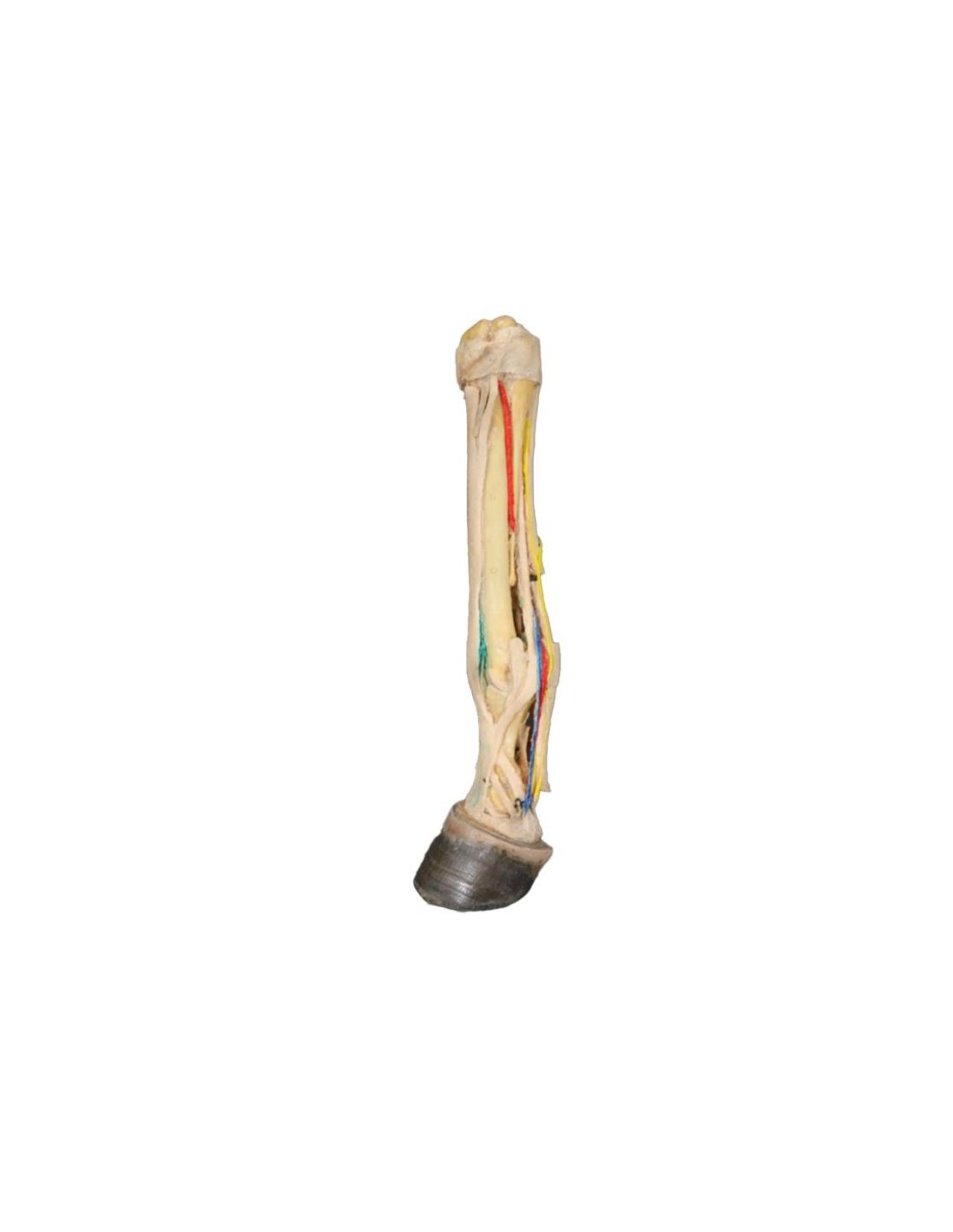
The Deep Palmar Dissection of the Horse’s Foot (VP10040) anatomical model is designed to provide an in-depth view of the internal structures of the palmar region of the equine foot. Thanks to this detailed dissection, it is possible to accurately study the muscles, tendons, blood vessels, nerves, and other anatomical components present in this crucial area of the horse. Ideal for anatomy courses, dissection labs, and research activities, the VP10040 is an essential tool for practical learning and specialized training in the veterinary field.
Key Features
Detailed and Realistic Anatomy
- Highly Accurate Reconstruction: The model precisely highlights the anatomical layers of the palmar region, clearly and systematically displaying tendons, ligaments, vessels, and nerves.
- Educational Insight: Deep dissection allows for the analysis of anatomical relationships and interconnections essential for understanding the biomechanics of the horse’s foot.
High-Quality Materials and Precision Finishing
- Guaranteed Durability: Manufactured with materials selected to ensure high durability, ideal for intensive use in academic and research settings.
- Attention to the Finest Details: Every element is carefully reproduced, offering a realistic and accurate study experience.
Wide Range of Educational and Research Applications
- Standard Educational Tool: Perfect for veterinary anatomy courses, dissection labs, and specialized seminars, it contributes to the acquisition of practical skills and the deepening of theoretical knowledge.
- Support for Clinical Research: Suitable for comparative studies and scientific investigations, the VP10040 facilitates detailed analysis of the internal structures of the equine foot.
Benefits of Use
Interactive and Safe Learning
Thanks to the Deep Palmar Dissection Model of the Horse’s Foot (VP10040), students and professionals can practice in a controlled environment, acquiring practical skills without the need to use live subjects. This ethical approach promotes safe and responsible learning.
A Valuable Investment for Institutions and Research Centers
This model represents a strategic investment for veterinary schools, universities, and research centers, contributing to the elevation of educational standards and clinical specialization thanks to its precision and reliability.
Technical Specifications
For more information on dimensions, materials used, and instructions for use, please consult the complete technical data sheet or contact our customer service for personalized assistance.
Buy Now
Don’t miss the opportunity to enrich your education or that of your students with the Deep Palmar Dissection
Model of the Horse’s Foot (VP10040)
.
Add to cart
and discover how this tool can revolutionize the study of veterinary anatomy, offering a detailed analysis of the structures fundamental to the horse’s health and performance.
What advantages does the Monash University anatomical dissection collection offer compared to plastic models or plastinated specimens?
- Each body replica has been carefully crafted from selected patient radiographic data or human cadaver specimens selected by a highly qualified team of anatomists at the Monash University Centre for Human Anatomy Education to illustrate a range of clinically important anatomical areas with a level of quality and accuracy that cannot be achieved with conventional anatomical models – this is real anatomy, not stylized.
- Each body replica has been rigorously checked by a team of highly qualified anatomists at the Center for Human Anatomy Education, Monash University, to ensure the anatomical accuracy of the final product.
- The body replicas are not real human tissue and are therefore not subject to any restrictions on transport, import, or use in educational institutions that do not hold an anatomy license. The Monash 3D Anatomy dissection series avoids these and other ethical issues that arise when dealing with plastinated human remains.
No reviews
Tap to zoom