Your cart
There are no more items in your cart


Sagittal Section of a Horse's Head VP10000
erler zimmer
EZ-VP10000
Out-of-Stock
€5,300.00
Tax included
Created using ultra-high-resolution 3D color printing.
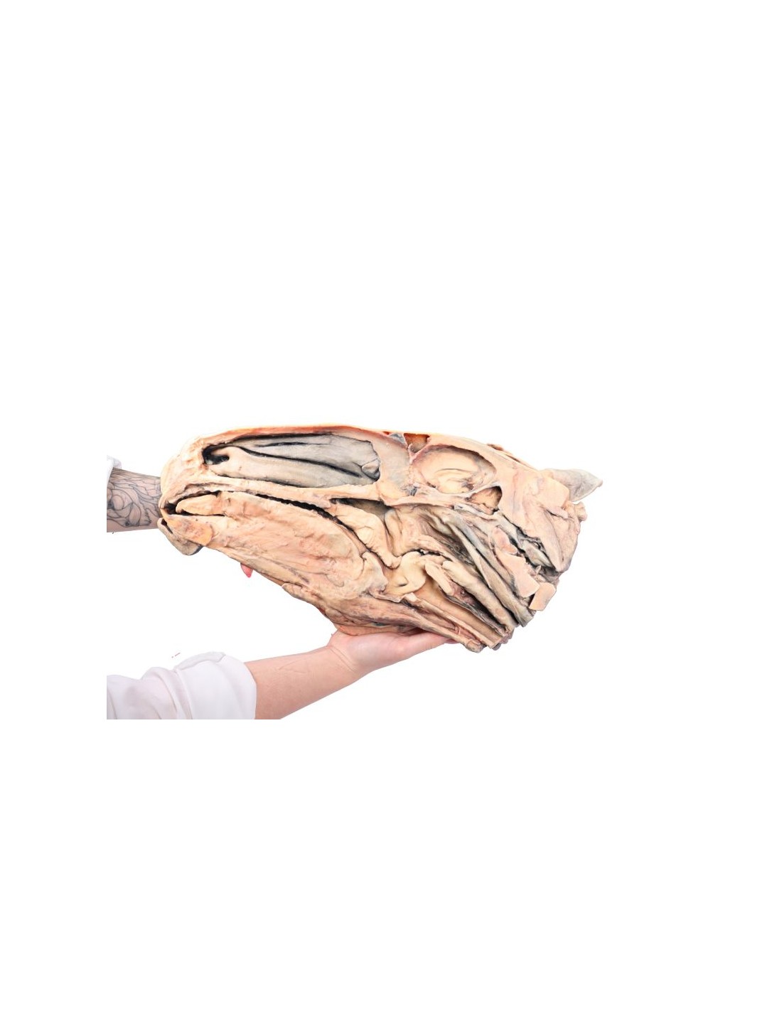
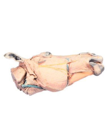
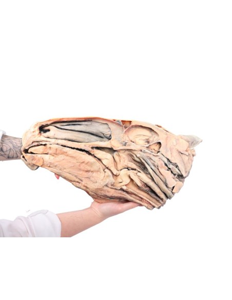
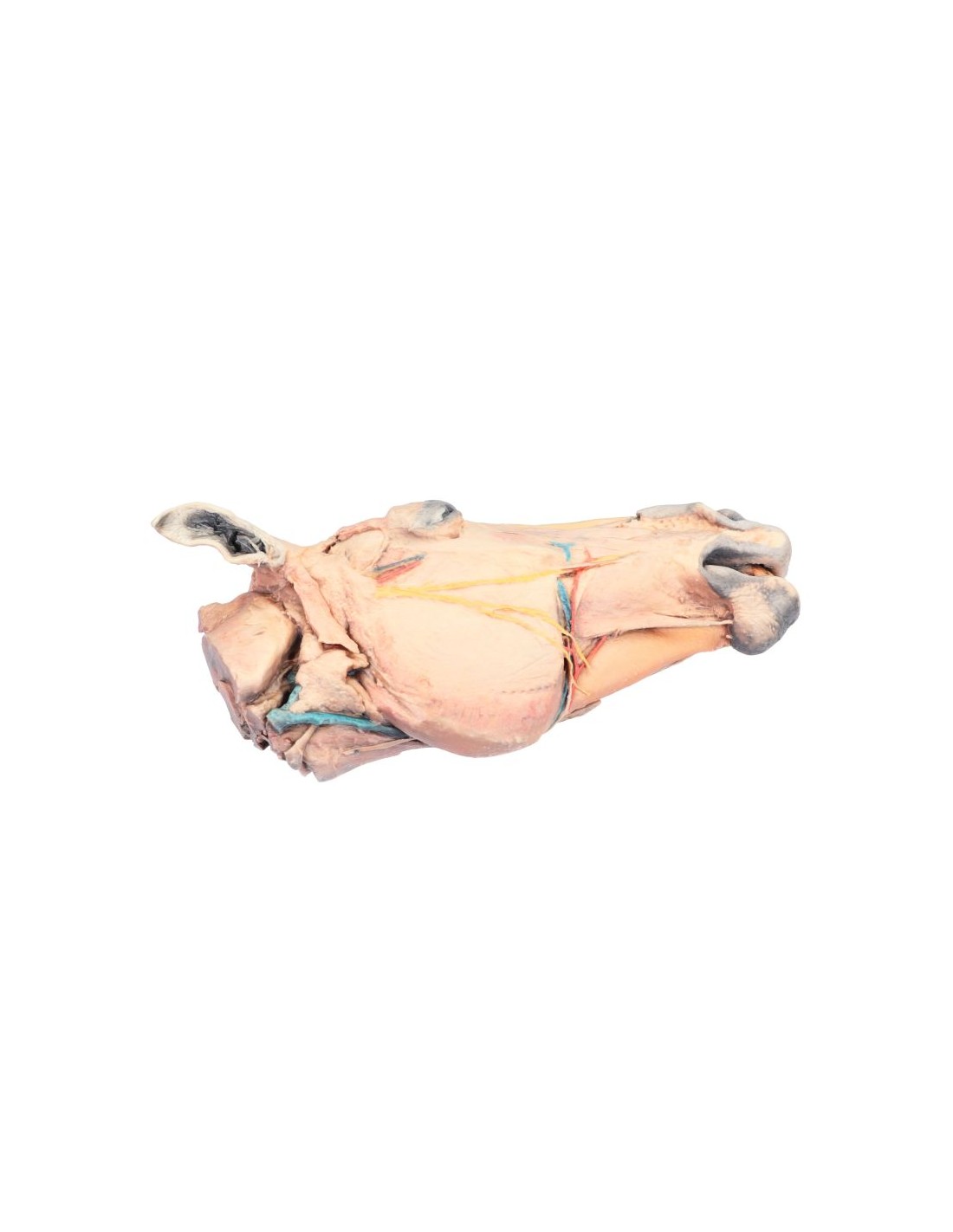
Sagittal Section of a Horse's Head VP10000
This model is part of the exclusive Monash 3D Anatomy series, a comprehensive collection of human dissections reproduced using high-resolution color 3D printing.
Product Description
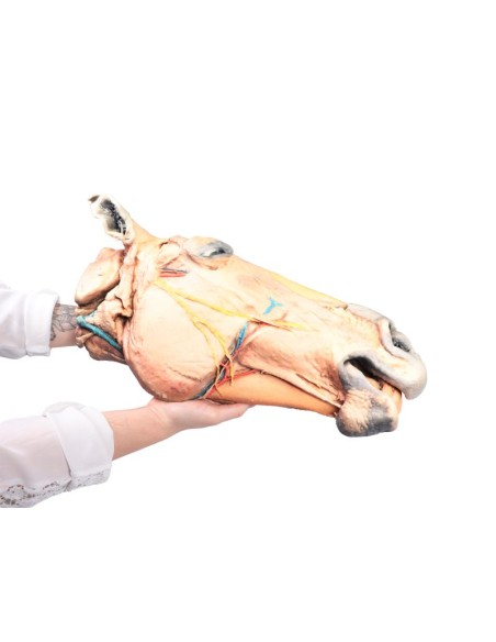
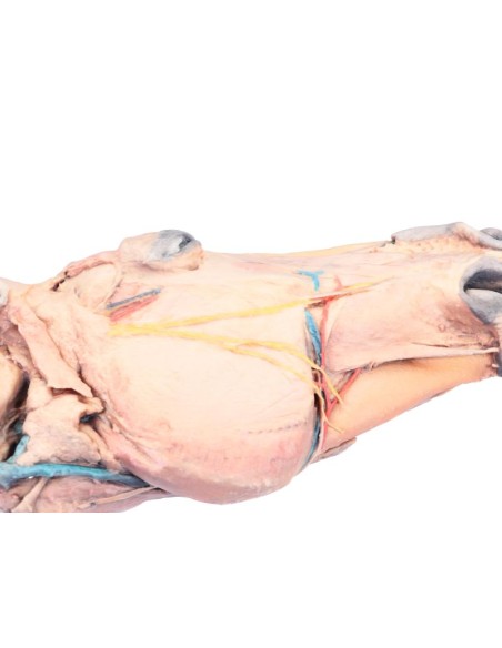
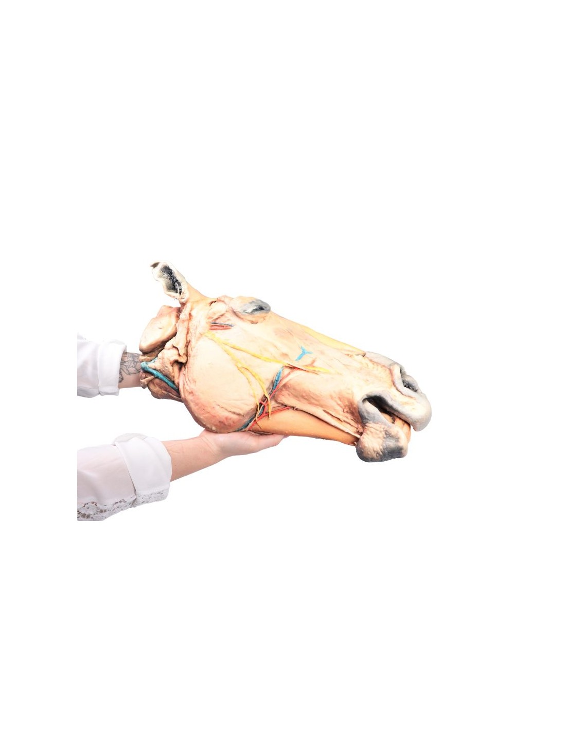
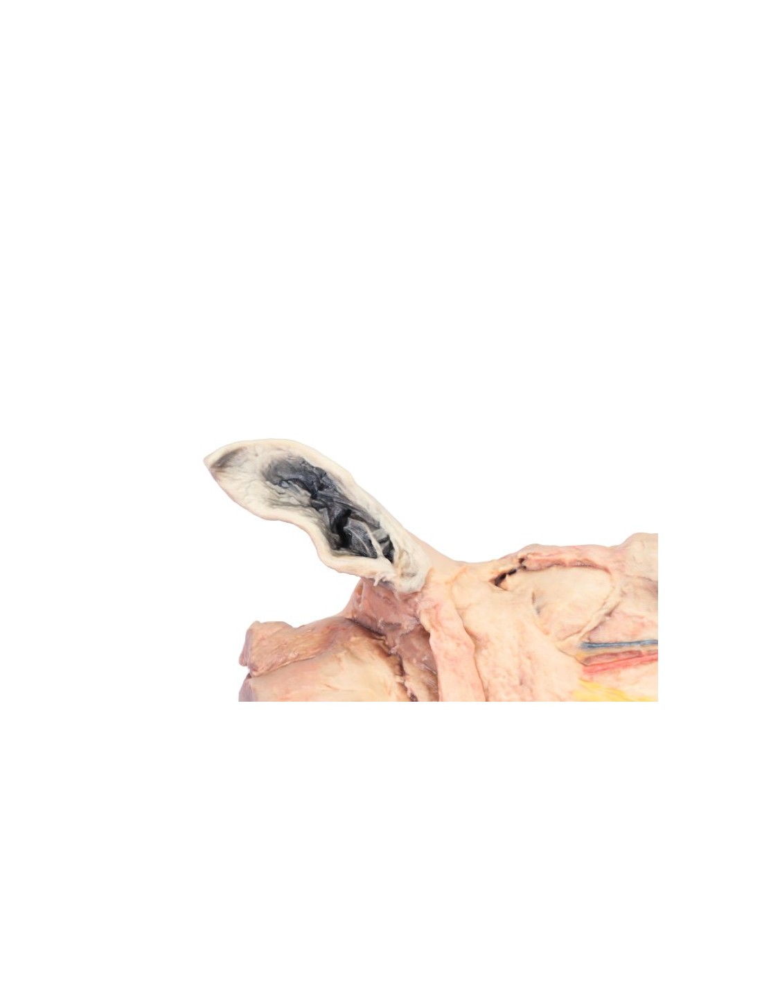
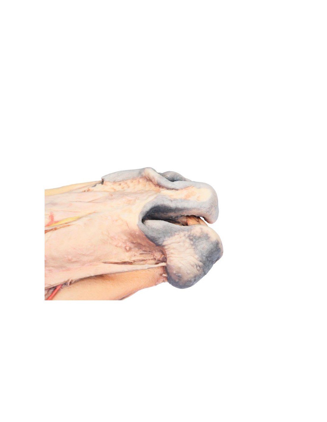
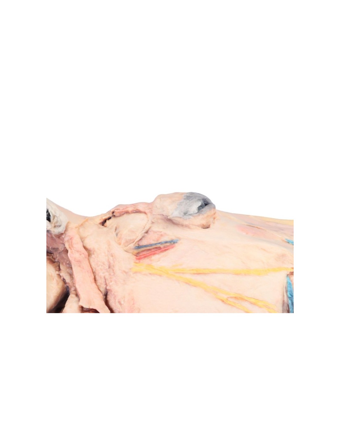
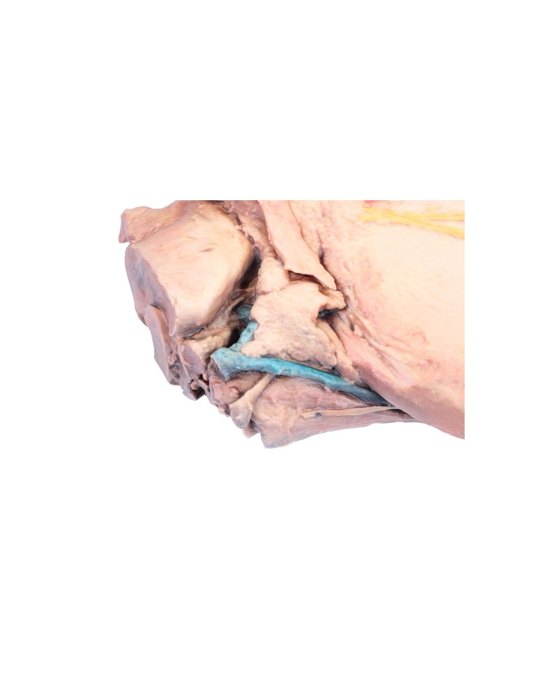
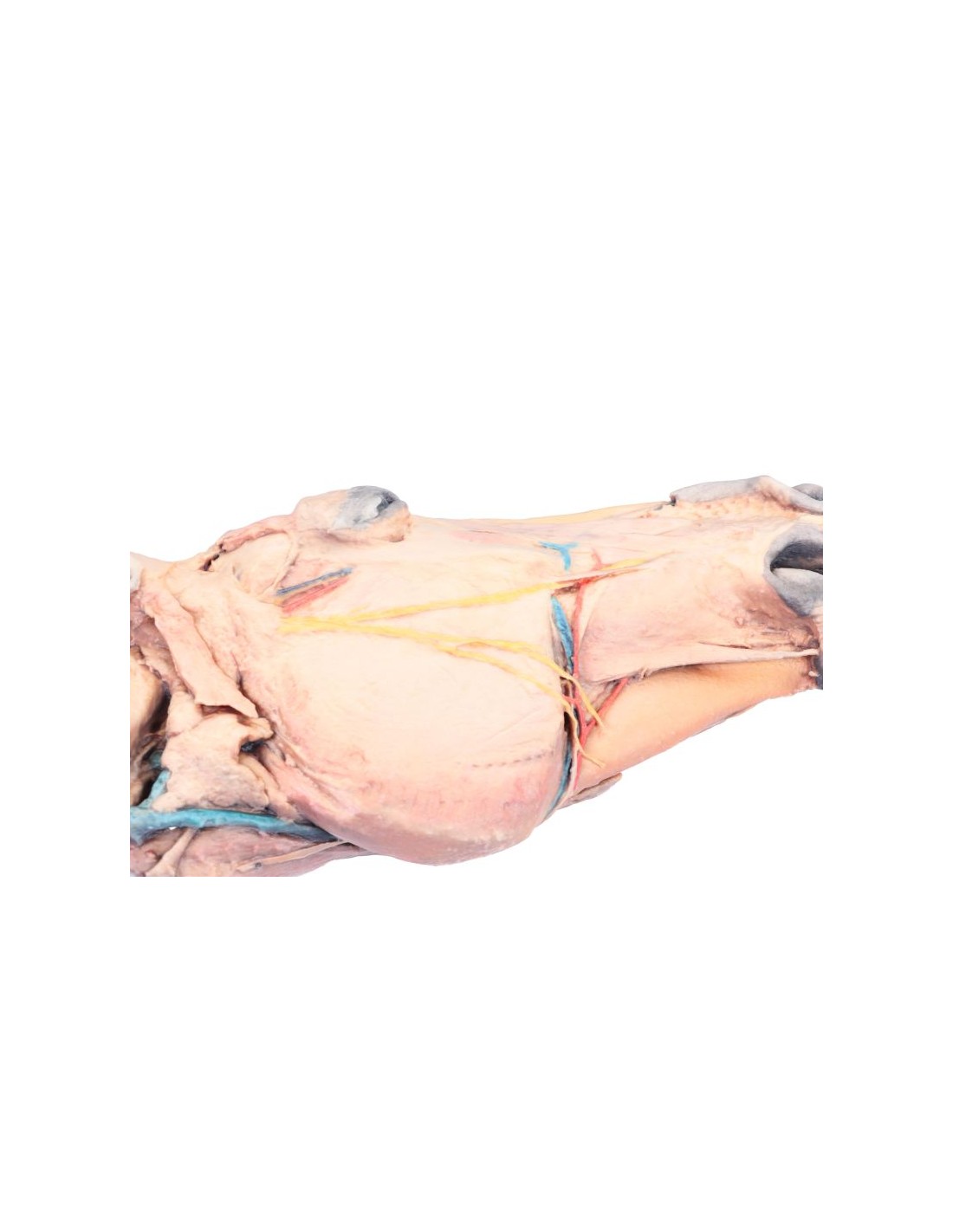
The Sagittal Section of the Horse’s Head anatomical model (VP10000) is an excellent educational tool for training and research in the veterinary field. This model, created as a precise sagittal section, allows for a detailed exploration of the internal structures of the equine skull, highlighting their anatomical and functional relationships. Ideal for anatomy courses, dissection labs, and comparative studies, the VP10000 offers an in-depth and realistic analysis of the morphology of the horse’s head.
Key Features
High-Precision Anatomical Representation
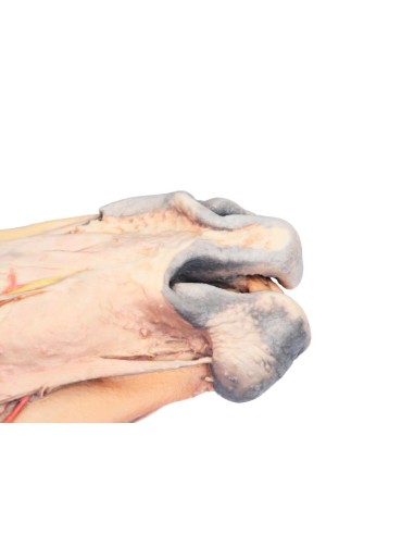
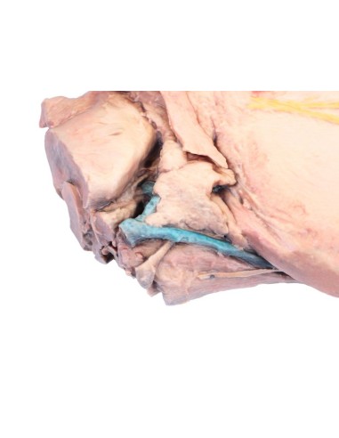
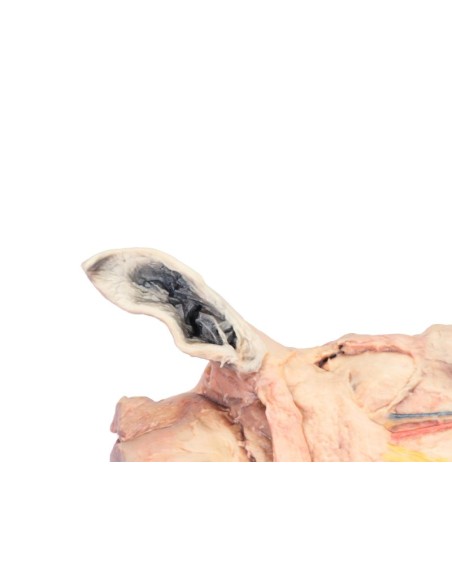
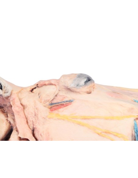
- Detail and Realism: The sagittal section accurately highlights the bone structures, muscles, nerves, and blood vessels within the equine skull, allowing for a comprehensive and detailed analysis.
- Study of Anatomical Relationships: The model facilitates understanding of the interconnections between various anatomical components, making learning interactive and in-depth.
High-Quality Materials and Finishes
- Durable Construction: Made with high-quality materials, the VP10000 is designed to ensure durability and resilience even in high-use environments such as veterinary schools and research centers.
- Attention to Detail: Every element of the model has been meticulously crafted to ensure a faithful and realistic representation of the complex equine anatomy.
Educational and Research Applications
- Advanced Educational Tool: Perfect for teaching and learning the morphology of the horse’s head, the model is ideal for dissection labs and veterinary anatomy courses.
- Support for Scientific Research: The VP10000 is ideal for research projects requiring detailed analysis of cranial structures, facilitating comparative studies and scientific investigations.
Benefits of Use
Interactive and Safe Learning
Using the Sagittal Section of the Horse’s Head model (VP10000), students and professionals can practice in a controlled environment, improving their understanding of anatomical structures in a practical and safe manner. This educational tool eliminates the need to use live subjects, ensuring an ethical and responsible approach to training.
Investment for Institutions and Research Centers
The VP10000 represents a strategic investment for universities, veterinary schools, and research centers, offering a high-quality educational tool that helps elevate the standard of teaching and clinical practice.
Technical Specifications
For more information regarding dimensions, materials used, and instructions for use, please consult the complete technical data sheet or contact our customer service for personalized assistance.
Buy Now
Don’t miss the opportunity to enrich your education or that of your students with the Sagittal Section of the Horse’s Head
model (VP10000)
.
Add it to your cart
and discover how this tool can revolutionize the approach to studying veterinary anatomy, offering a detailed and realistic view of equine cranial structures.
What advantages does the Monash University anatomical dissection collection offer compared to plastic models or plastinated specimens?
- Each body replica has been carefully crafted from selected patient radiographic data or human cadaver specimens selected by a highly qualified team of anatomists at the Monash University Centre for Human Anatomy Education to illustrate a range of clinically important anatomical areas with a level of quality and accuracy that cannot be achieved with conventional anatomical models – this is real anatomy, not stylized.
- Each body replica has been rigorously checked by a team of highly qualified anatomists at the Center for Human Anatomy Education, Monash University, to ensure the anatomical accuracy of the final product.
- The body replicas are not real human tissue and are therefore not subject to any restrictions on transport, import, or use in educational institutions that do not hold an anatomy license. The Monash 3D Anatomy dissection series avoids these and other ethical issues that arise when dealing with plastinated human remains.
No reviews
Tap to zoom