
Your cart
There are no more items in your cart


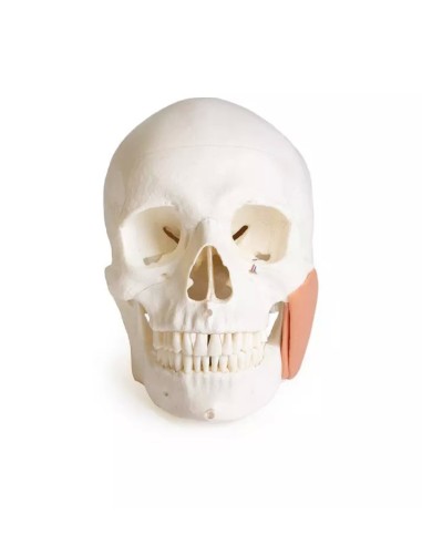
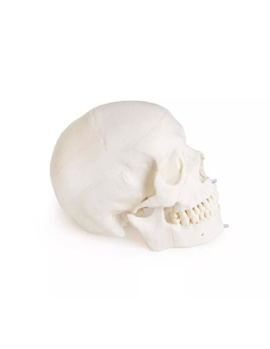

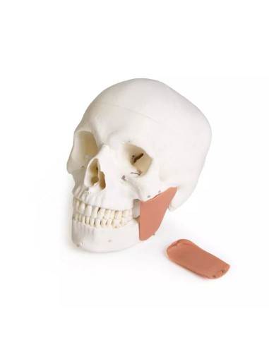
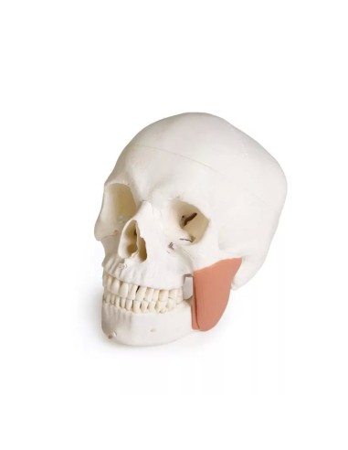




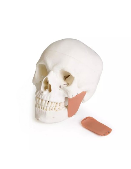


Classic, life-size anatomical skull model for dentistry
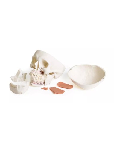
8-Part Skull Model for Dentistry with CMD Syndrome, EZ-4860:
An 8-part anatomical model of the human skull, specifically designed for the study of dentistry, maxillofacial surgery, and craniomandibular dysfunction (CMD).
In addition to reproducing the main anatomical structures of the skull with a high level of detail, the model focuses on the teeth, maxilla, mandible, temporomandibular joint, and masticatory muscles, allowing for the study of both normal anatomy and certain abnormalities typical of craniomandibular dysfunction.
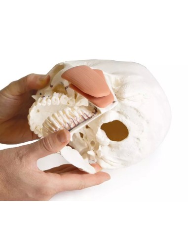
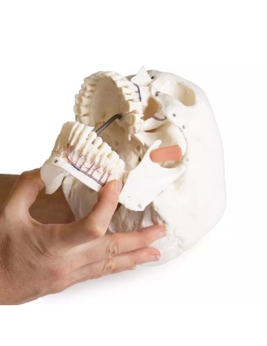
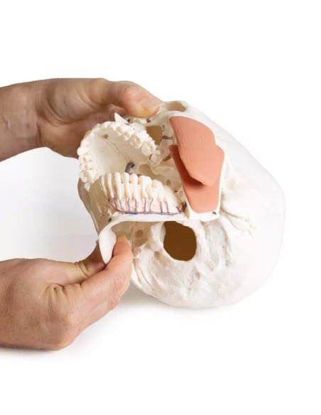
The front portion of the maxilla and mandible is removable, allowing for direct observation of the teeth and their roots. Nerves and blood vessels are also depicted on one side, while the opening of the maxilla allows for a clear view of the maxillary sinuses as well.
A distinctive feature of this version is the depiction of craniomandibular dysfunction (CMD), a condition characterized by a functional alteration in the interaction between the temporomandibular joints, the masticatory muscles, and the dentition.
The model highlights several characteristic findings, including:
- tooth enamel wear resulting from bruxism;
- wear of the articular disc of the temporomandibular joint;
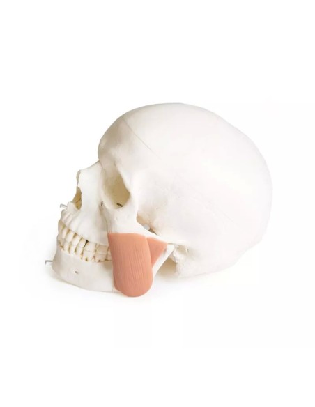
- a detailed representation of the main portions of the masseter muscle involved in masticatory function.
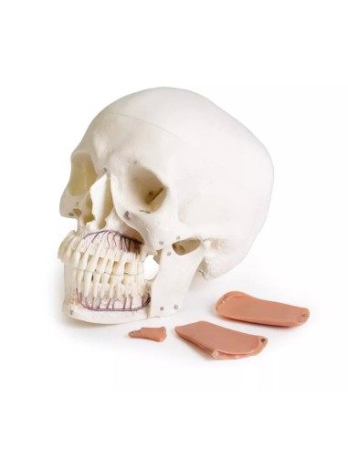
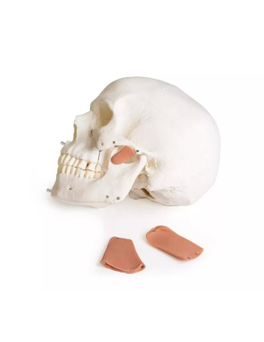
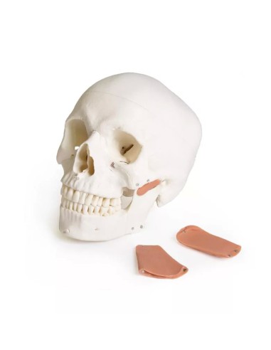
The three portions of the masseter muscle are depicted:
- Masseter muscle, superficial portion
- M. masseter, pars profunda
- M. masseter, pars coronoidea
All three muscle components are removable, allowing for a clearer view of their relationships with the mandible and surrounding structures.
Key Features
- 8-part anatomical model of the human skull
- Specifically developed for dentistry and maxillofacial surgery
- Depiction of craniomandibular dysfunction (CMD)
- Removable upper and lower jaws at the front
- Teeth with visible anatomical roots
- Unilateral depiction of nerves and blood vessels
- Visualization of the maxillary sinuses
- Depiction of dental wear caused by bruxism
- Depiction of wear on the temporomandibular joint disc
- Masseter muscle depicted in its superficial, deep, and coronoid portions
- All three muscle sections are removable
The model is particularly suitable for students and faculty in dentistry, medicine, and anatomy, as well as for dentists, maxillofacial surgeons, orthodontists, gnathologists, and professionals who treat temporomandibular joint disorders.
Thanks to the ability to disassemble various structures, it can be used both as a tool for anatomical study and for clinical training and explaining to patients issues related to occlusion, bruxism, and craniomandibular dysfunction.
Why Buy It
Erler Zimmer anatomical models are the most widely used teaching aids for the study of human anatomy in schools and universities. High quality, attention to detail, and excellent value for money.
Ideal for teaching, explaining conditions to patients, and medical-scientific training.
Many doctors and professionals purchase these educational anatomical models to highlight key points on topics such as the skeleton, the human musculature, joints, and related conditions (rheumatism, osteoarthritis, bursitis, synovitis, tendinitis, cervical pain, and sciatica).
They can also be used as decorative accents to personalize your medical office.
- Height
- 19
- Width
- 18
- Depth
- 12
- Weight
- 0,7
No reviews
Tap to zoom
