
Your cart
There are no more items in your cart




Complete Anatomical Dissection of a Cat – Erler Zimmer VP12000
erler zimmer
EZ-VP12000
€4,450.00
Tax included
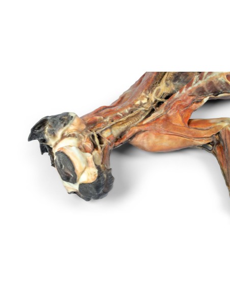
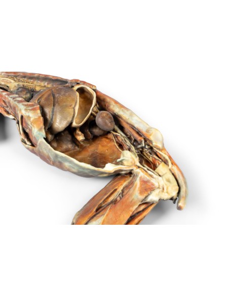
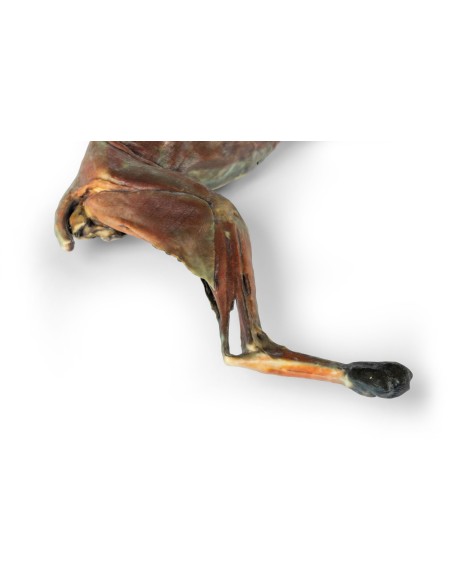
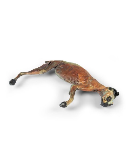
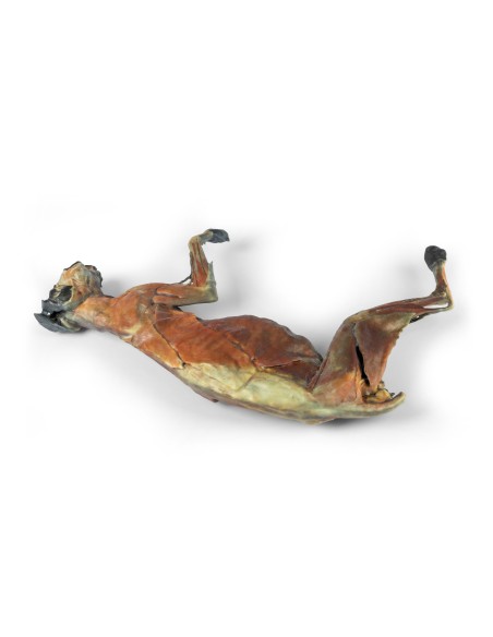
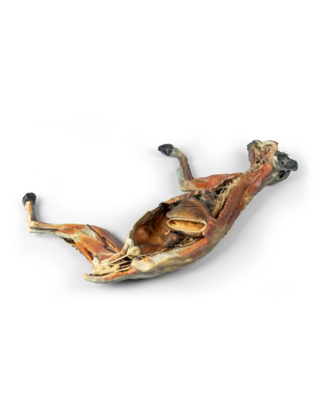
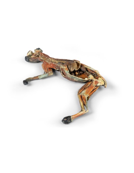
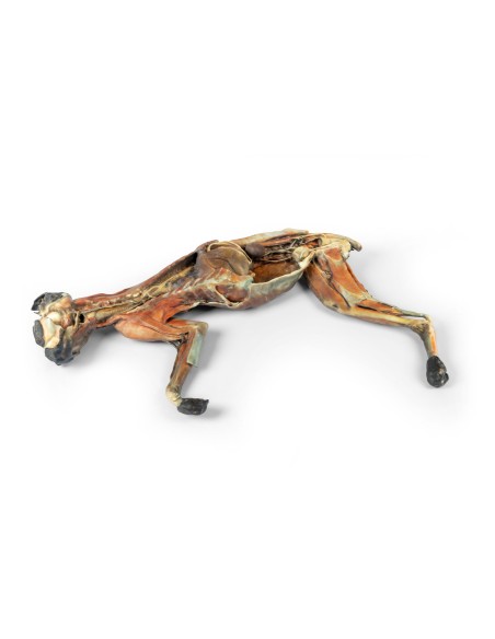
A 3D anatomical model of the entire cat’s body with two complementary dissections. One side shows the head, neck, and thoracic, abdominal, and pelvic cavities, while the opposite side highlights the superficial musculature and the relationships between the trunk and the limbs.
Complete Anatomical Dissection of the Cat – Erler Zimmer VP12000
The Complete Anatomical Dissection of the Cat – Erler Zimmer VP12000 reproduces a dissection of the entire cat’s body and offers two complementary anatomical views, designed to allow the study of both the topography of the internal organs and the superficial organization of the muscular system in a single specimen.
One side of the model depicts the dissection of the head and neck and the opening of the thoracic, abdominal, and pelvic cavities. This configuration allows for the observation of numerous organs and anatomical structures while maintaining their relative positions within the body, facilitating an understanding of topographic anatomy as a whole.
The continuity of the specimen, from the cranial region to the pelvis, allows structures belonging to different systems to be related to one another without losing reference to their natural positions. The model is therefore particularly useful when the study focuses not on a single organ but on the overall organization of the cat’s anatomy.
On the opposite side, a superficial dissection of the musculature is visible. This second view offers a functional perspective on the musculoskeletal system and allows for a better understanding of the connection between the musculature of the trunk and that of the forelimbs and hindlimbs.
The ability to compare, within the same model, the arrangement of the internal organs with that of the superficial musculature represents one of the most interesting educational aspects of the VP12000. Students can quickly shift from studying visceral anatomy to that of the musculoskeletal system, while maintaining a three-dimensional and coherent view of the entire animal.
The model is particularly well-suited for university courses in Veterinary Medicine, for comparative and topographic anatomy exercises, and for laboratories where it is useful to have a complete, stable dissection that can be easily observed by multiple students.
No reviews
Tap to zoom
