Your cart
There are no more items in your cart
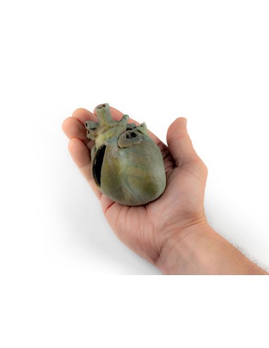


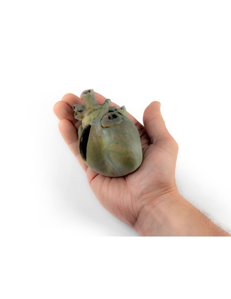
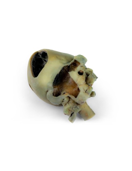
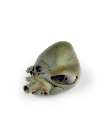
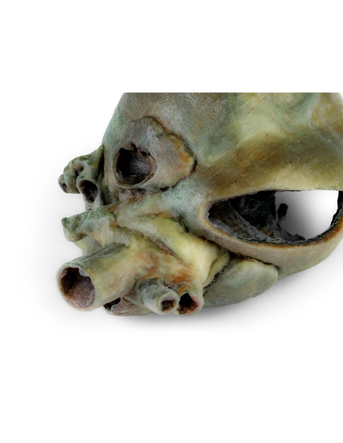
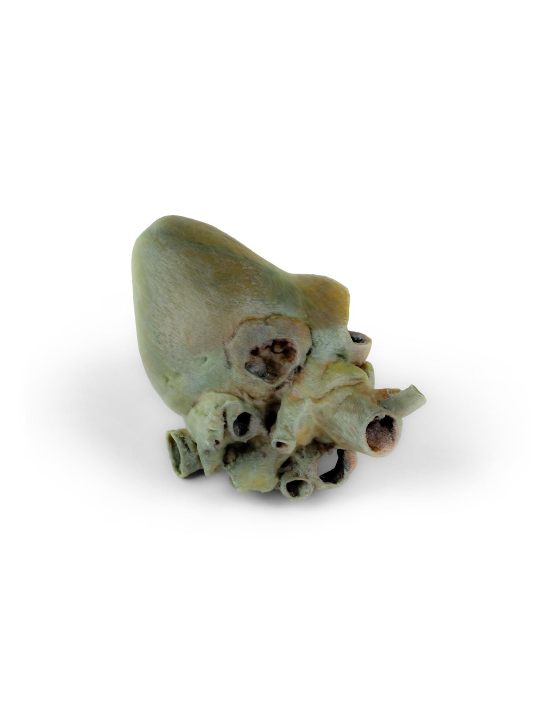
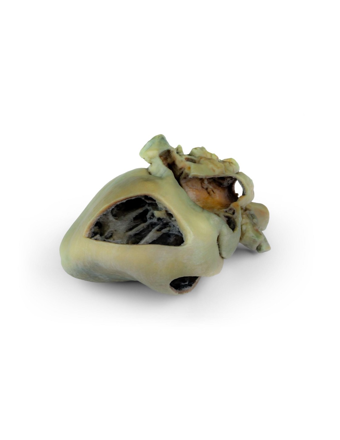
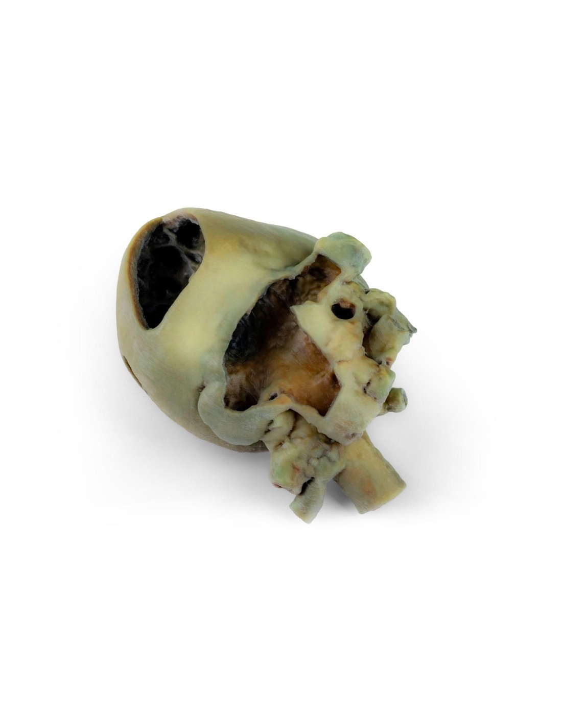
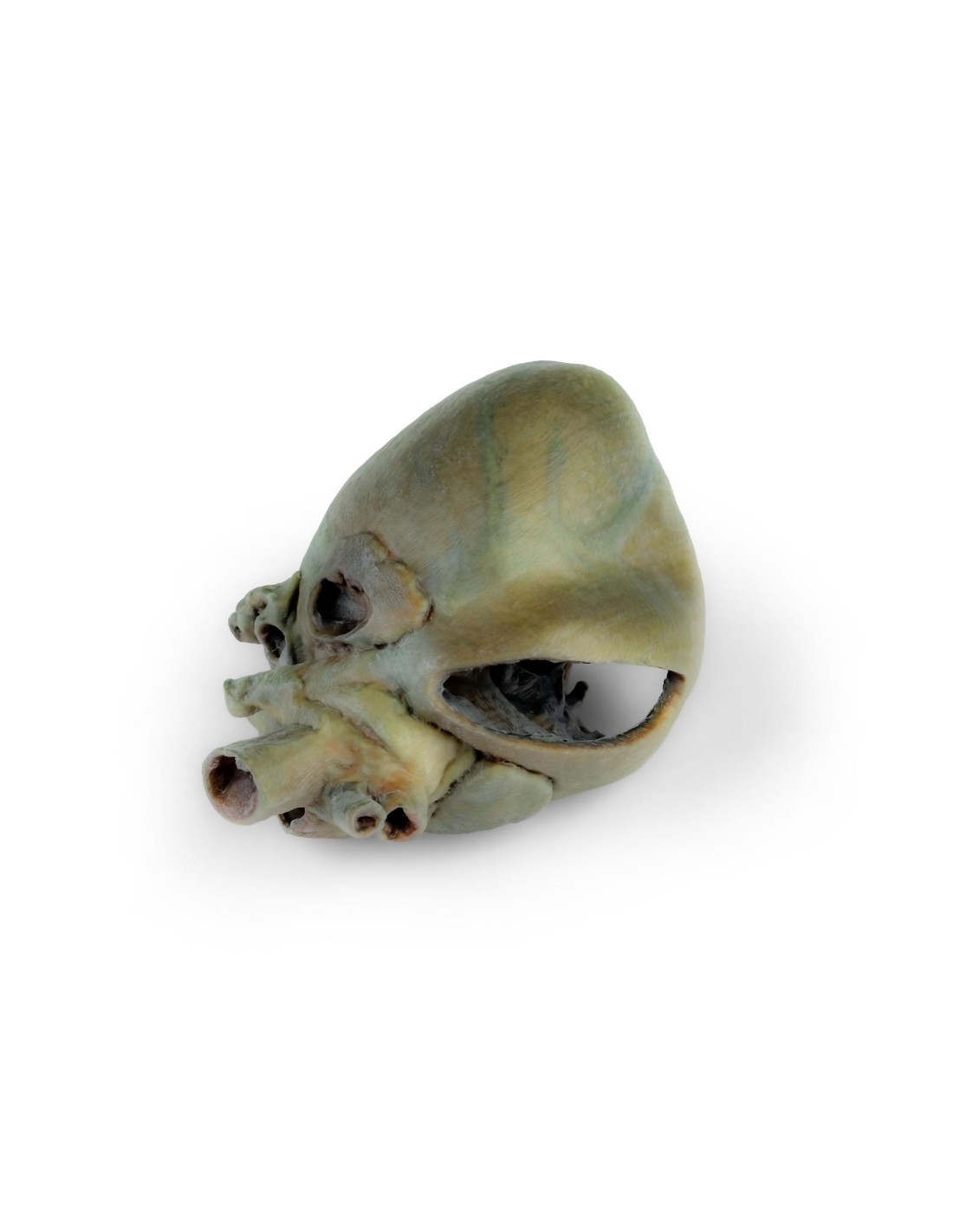
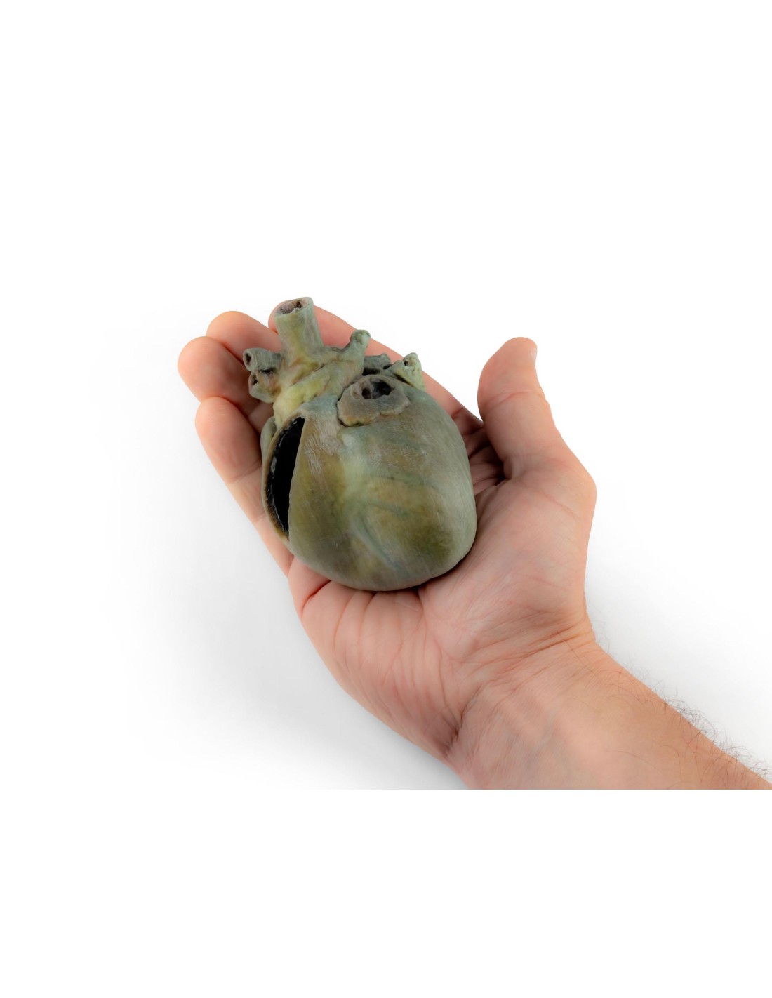
Dog Heart with Anatomical Dissection – Erler Zimmer VP9030
erler zimmer
EZ-VP9030
€650.00
Tax included
A 3D anatomical model of a dog’s thoracic cavity, viewed from the left side. Removal of the left lung exposes the heart, the great vessels, the esophagus, the diaphragm, and the major structures of the thoracic region and the cranial abdomen.
Dog Heart with Anatomical Dissection – Erler Zimmer VP9030
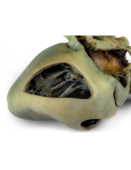
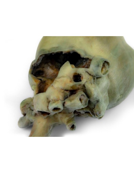
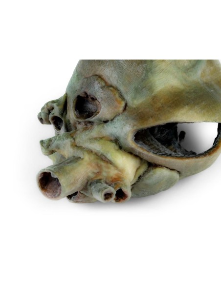
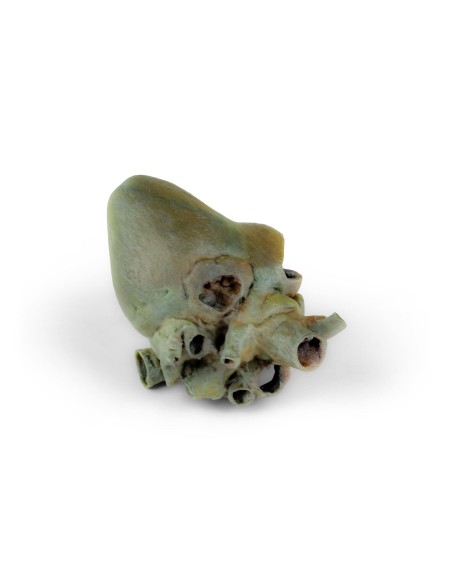
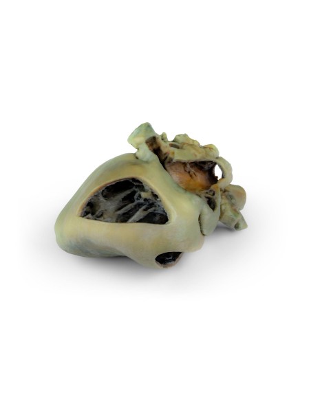
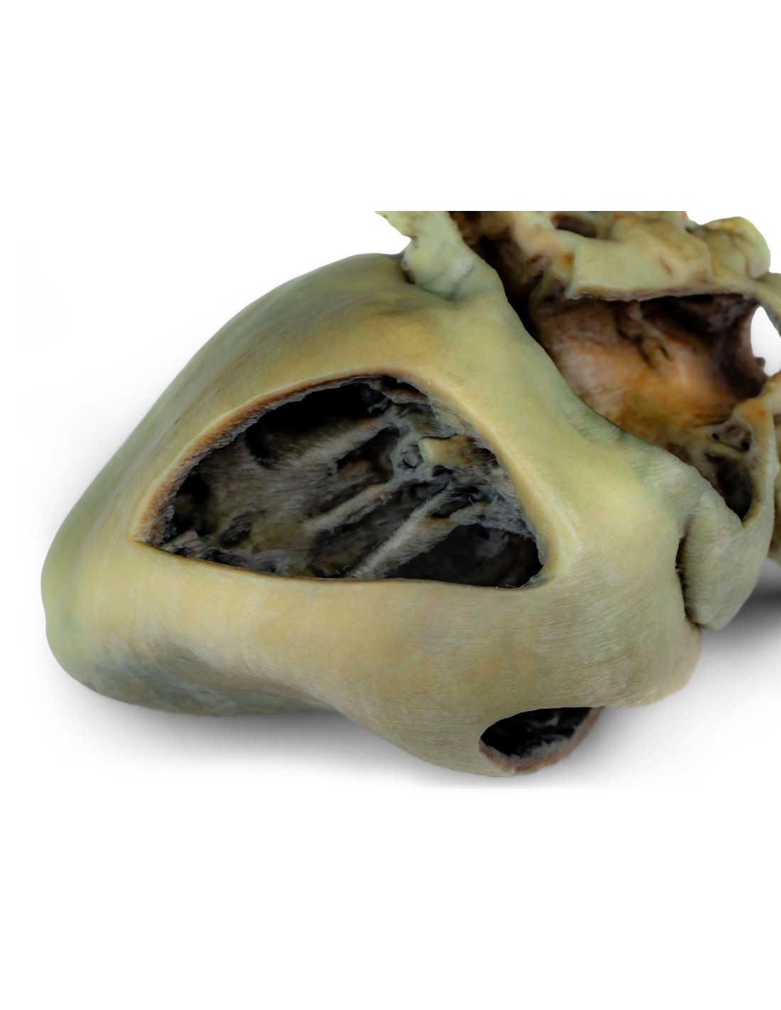
The Erler Zimmer VP9030 model reproduces a dog’s heart that has undergone anatomical dissection, with portions of the heart walls opened to allow direct observation of the internal chambers and the major valve structures.
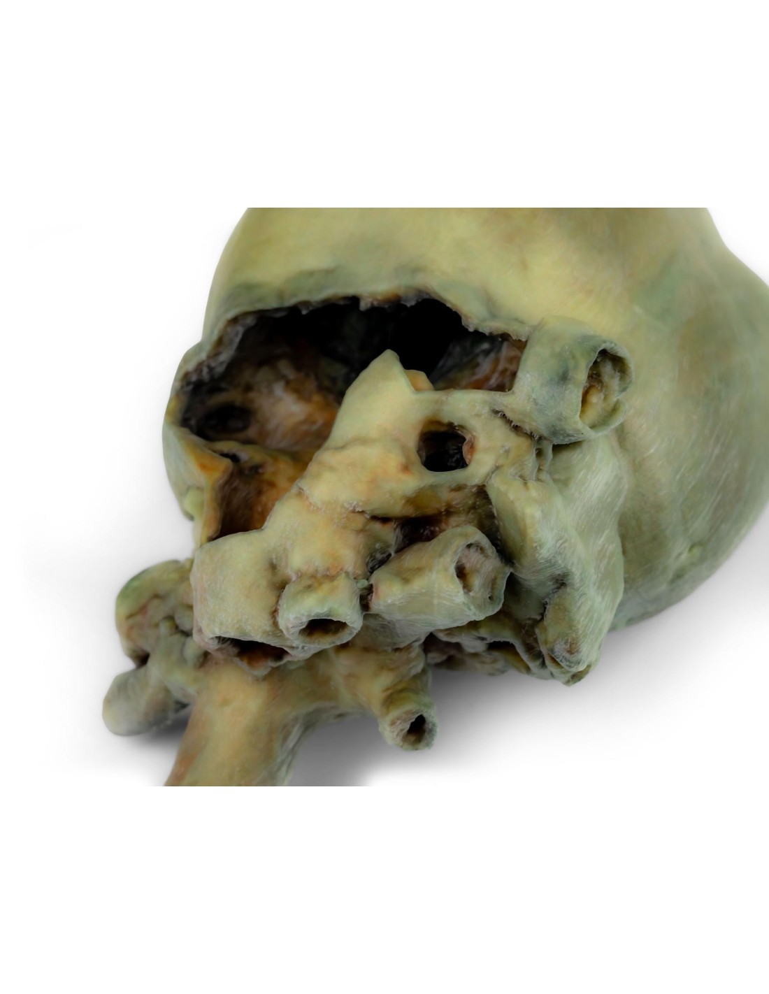
The outer surface of the heart retains the main anatomical landmarks, allowing users to identify the atrial and auricular regions and to trace the course of the interventricular and coronary sulci. At the base of the heart, the ascending aorta and its relationship to the pulmonary trunk are clearly visible, along with the openings of the cranial and caudal vena cavae into the right atrium.
The specimen also allows for observation of the bifurcation of the pulmonary trunk into the pulmonary arteries and the opening of the pulmonary veins into the left atrium. These features make the model particularly useful for understanding the arrangement of the major vessels entering and exiting the heart and their relationships with the cardiac chambers.
A portion of the right atrium wall has been removed to expose the right atrioventricular valve, or tricuspid valve. The chordae tendineae and papillary muscles—structures essential for understanding the functioning of the valvular system—are also clearly visible.
Inside the right ventricle, the septomarginal trabecula, also known as the moderator band, is visible, along with the tract leading from the ventricle to the pulmonary trunk. The wall of the left atrium has also been opened, allowing for direct observation of its lumen and enabling the atrial cavity to be correlated with the openings of the pulmonary veins.
The combination of external anatomy and dissection of the internal chambers makes the VP9030 particularly well-suited for studying canine cardiac morphology. The model provides a three-dimensional understanding of the arrangement of the heart chambers, valves, and great vessels, offering effective educational support both during lectures and in practical exercises.
It is recommended for university courses in Veterinary Medicine, for teaching cardiovascular anatomy and physiology, and for all educational activities in which it is useful to directly observe the relationships between the external and internal structures of the canine heart.
No reviews
Tap to zoom