Your cart
There are no more items in your cart


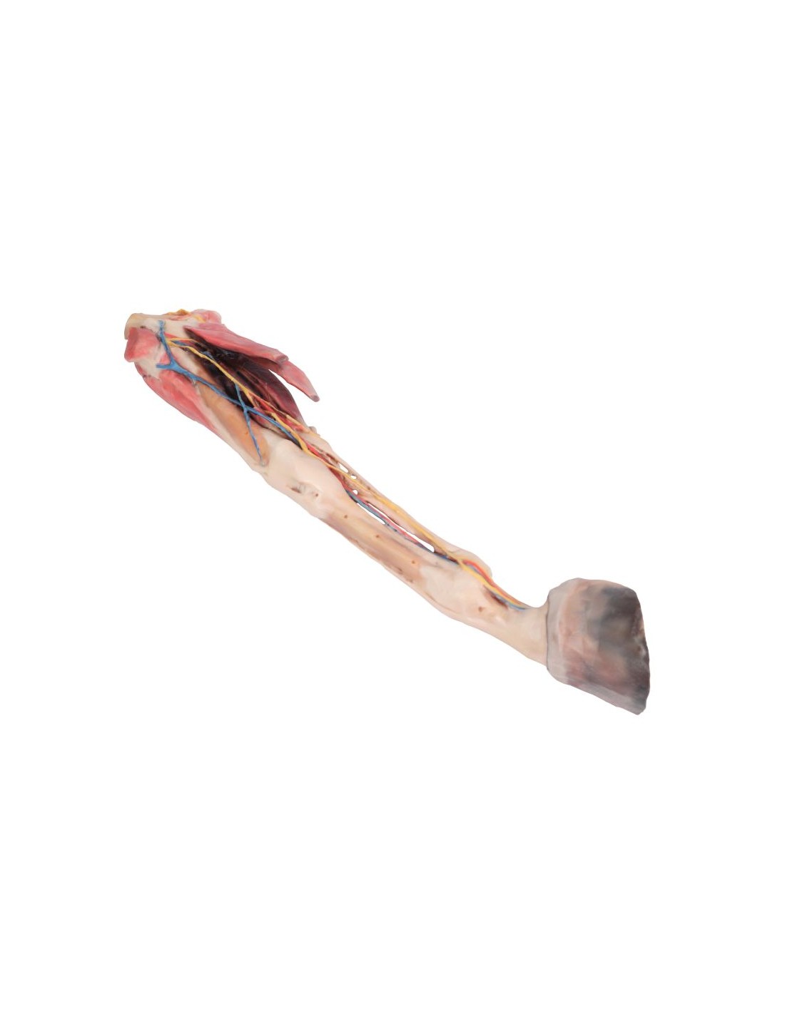
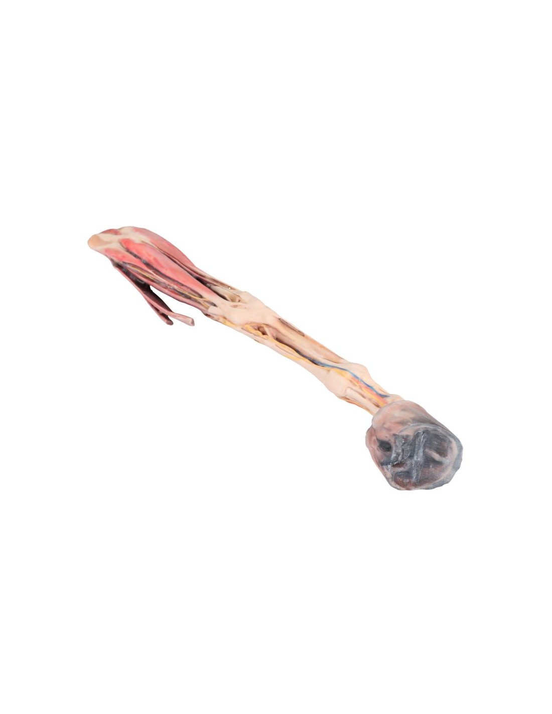
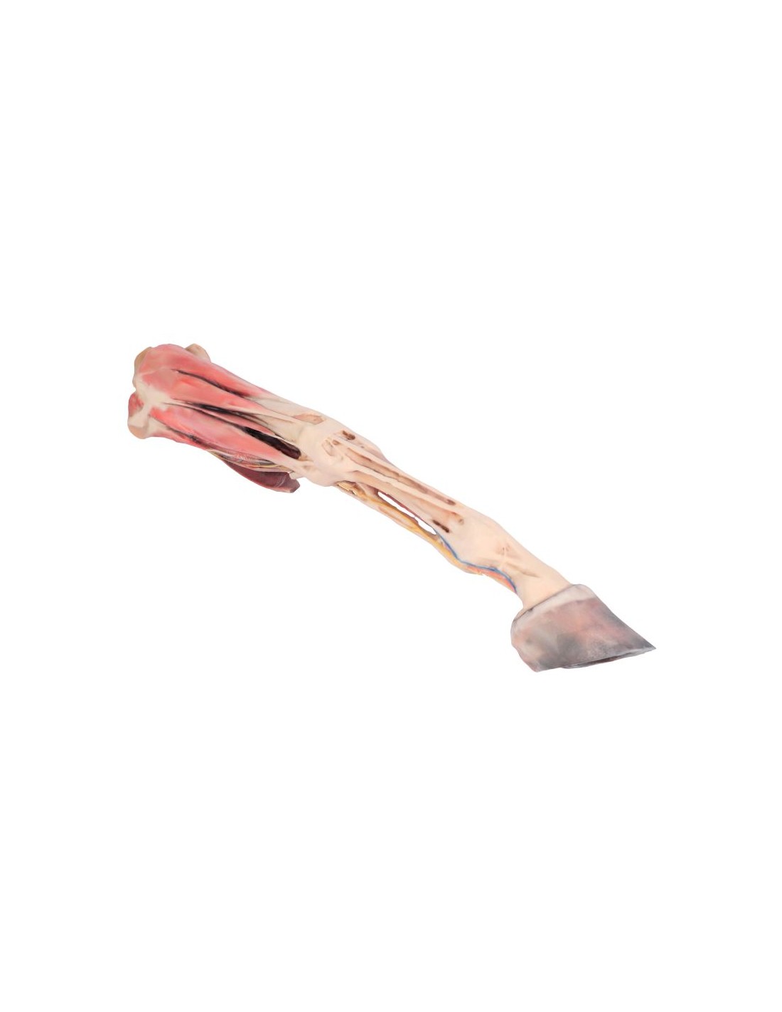
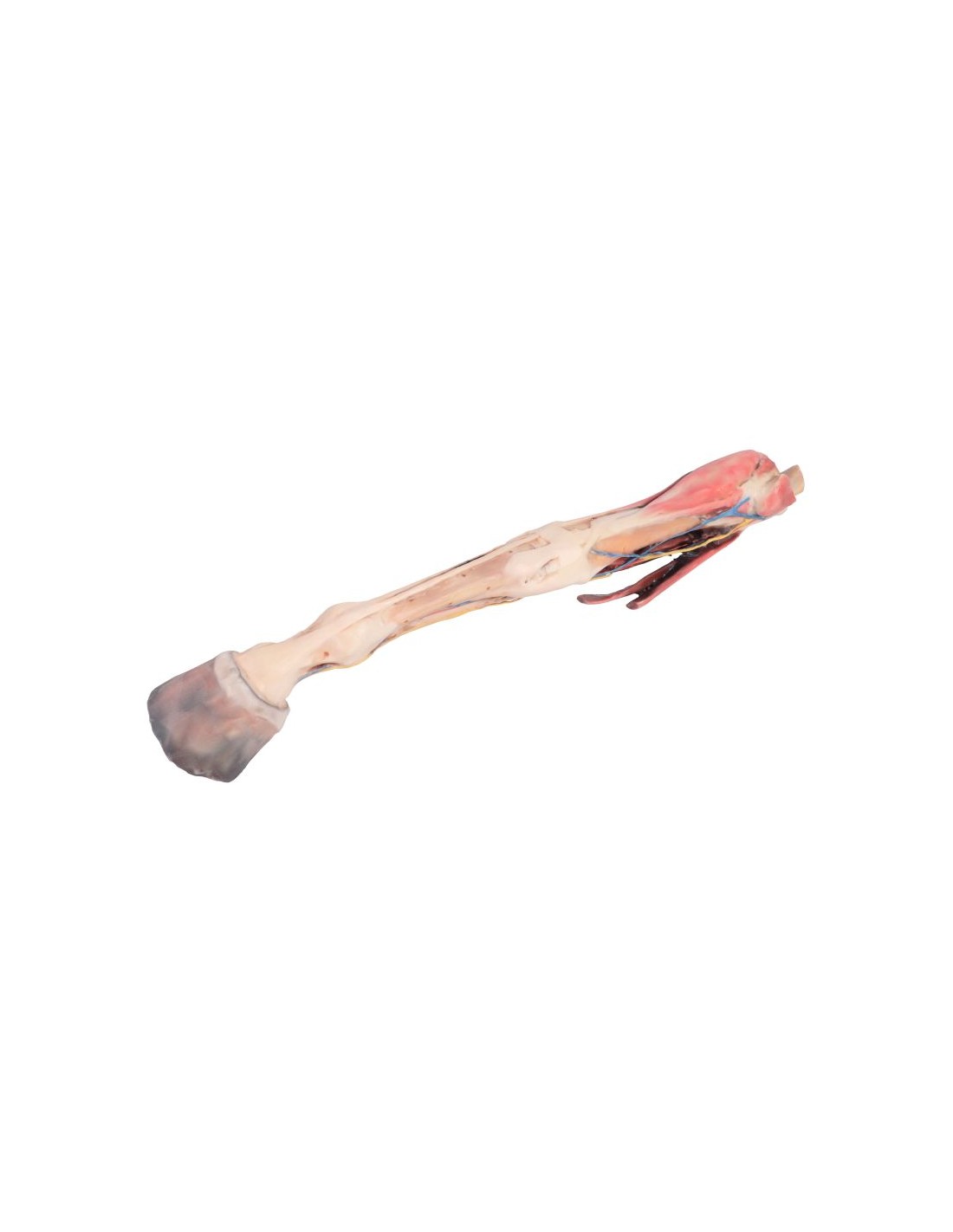
The Horse's Forequarter – Muscles, Tendons, Ligaments, Blood Vessels, and Distal Nerves VP10010
erler zimmer
EZ-VP10010
Out-of-Stock
€2,800.00
Tax included
Created using ultra-high-resolution 3D color printing.
Horse's Fore肢 – Muscles, Tendons, Ligaments, Vessels, and Distal Nerves Down to the Knee Joint, Scale VP10010
This model is part of the exclusive Monash 3D Anatomy series, a comprehensive collection of human dissections reproduced using high-resolution color 3D printing.
Product Description
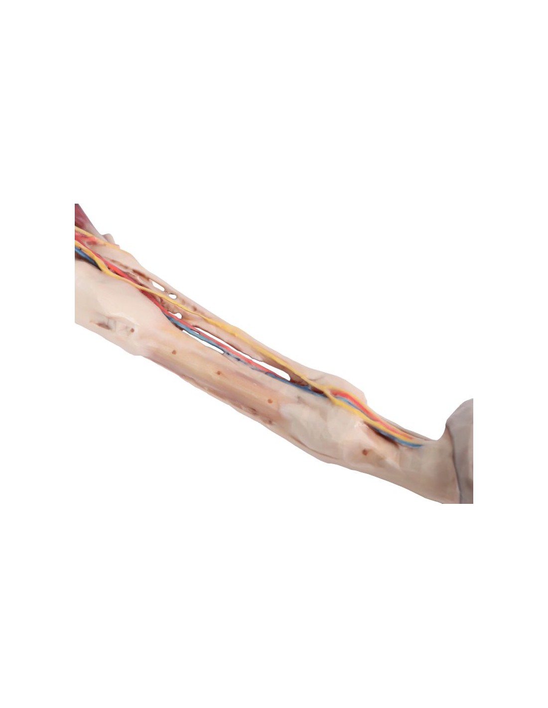
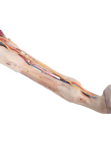
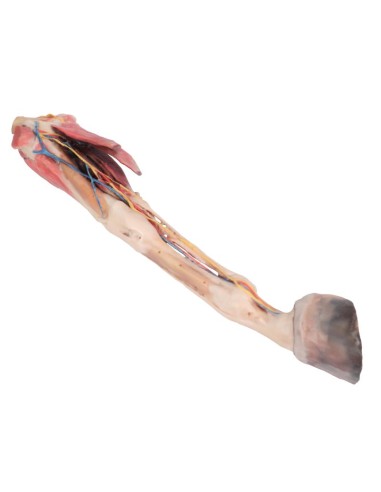
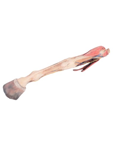
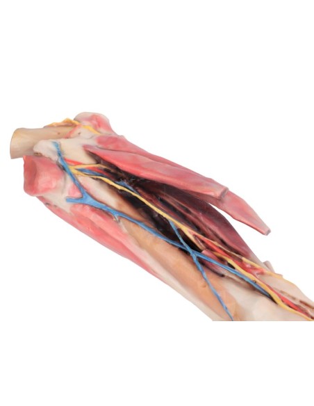
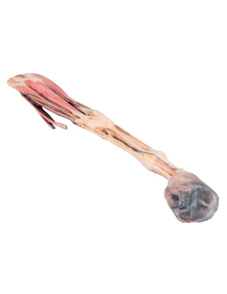
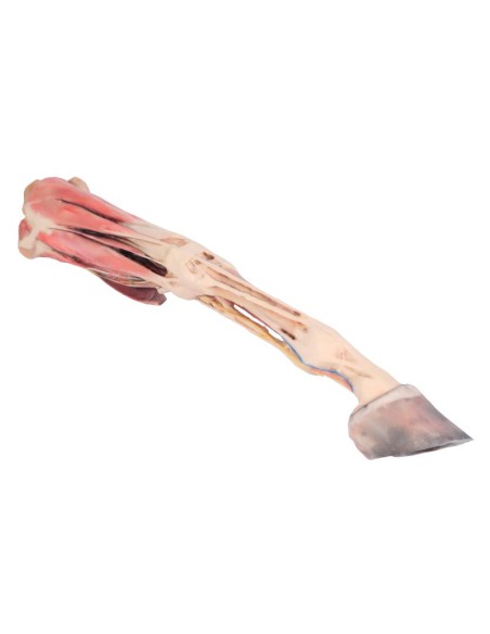
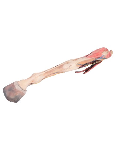
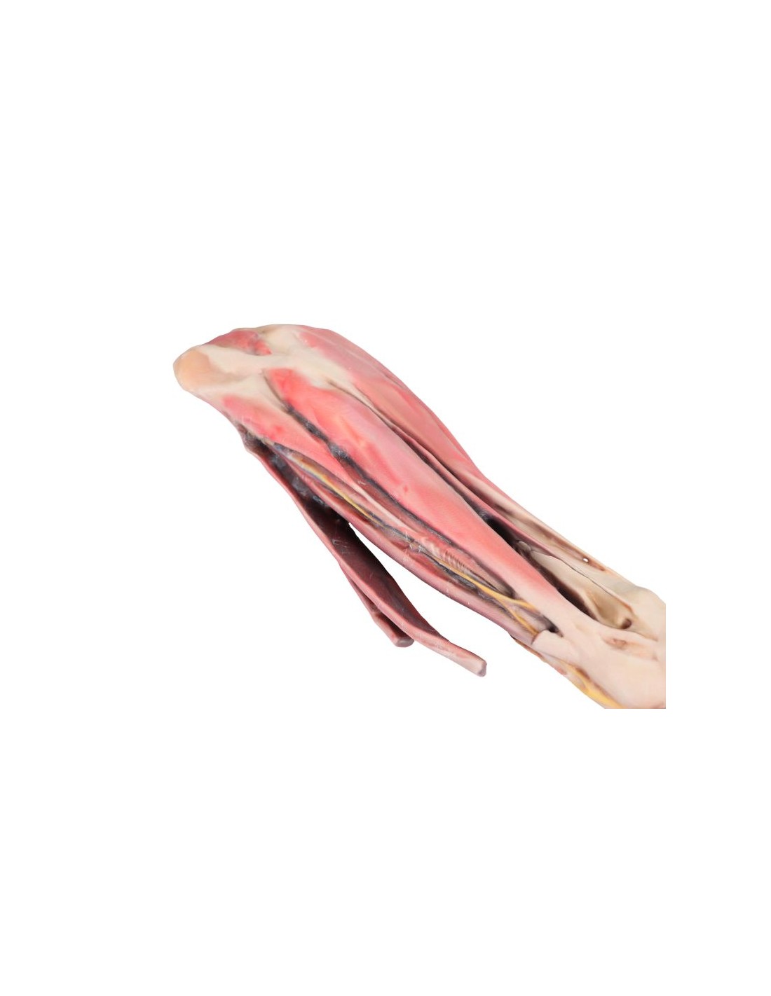
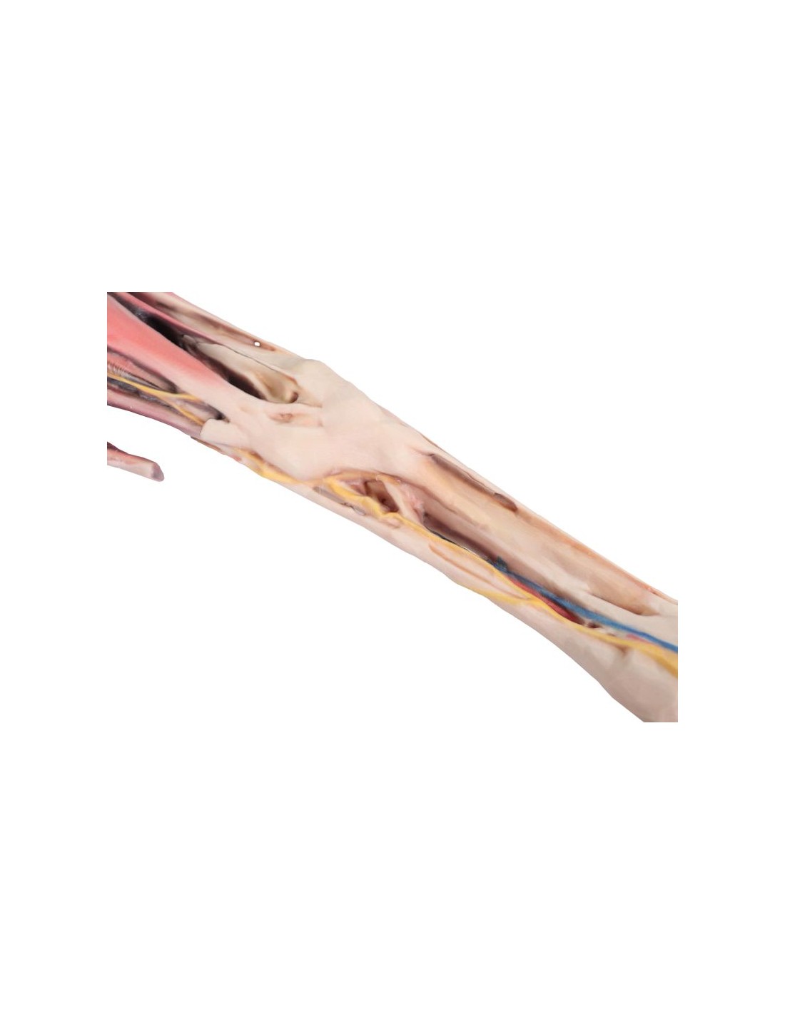
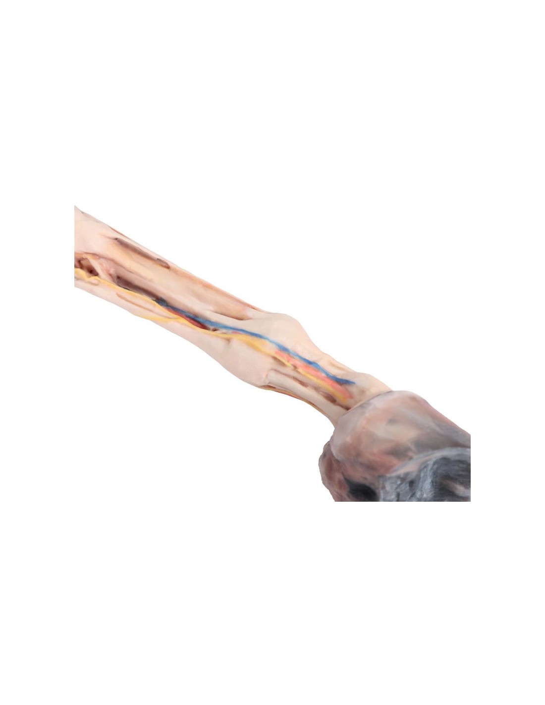
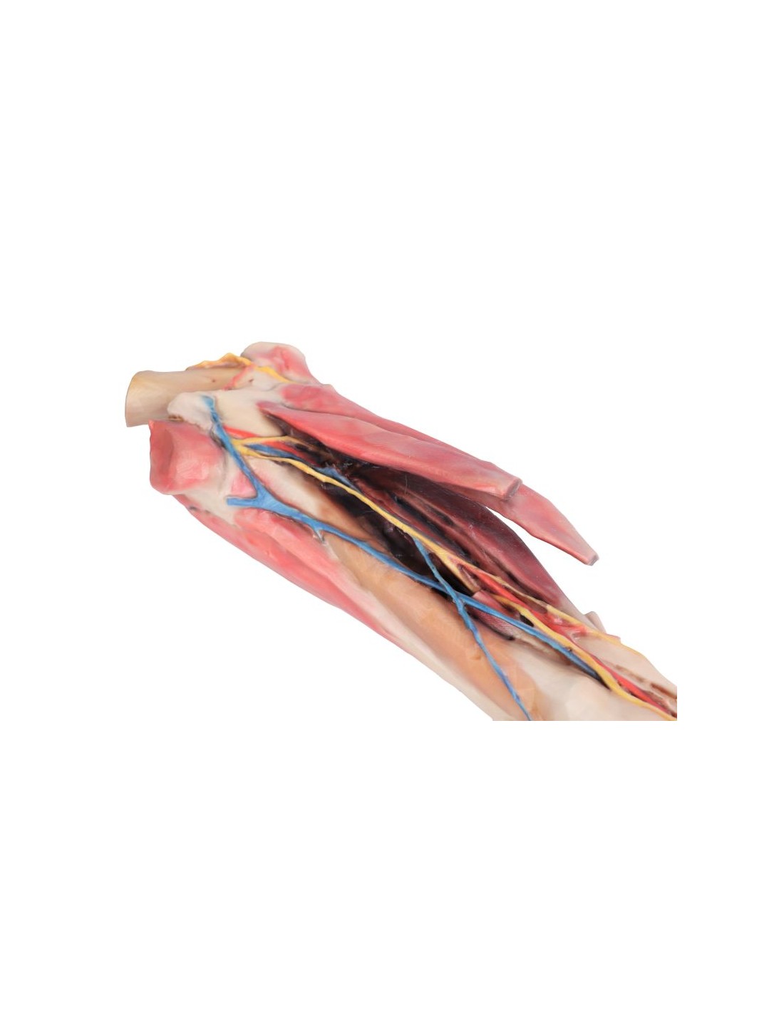
The Horse Fore肢 – Muscles, Tendons, Ligaments, Vessels, and Distal Nerves up to the Knee Joint (Scale 1:3) anatomical model is a high-quality educational tool, created at a 1:3 scale to provide an enlarged and detailed view of the anatomy of the horse’s forelimbs. Ideal for veterinary anatomy courses, dissection labs, and comparative studies, this model allows for in-depth examination of the muscular, tendinous, ligamentous, vascular, and nervous structures, facilitating practical and interactive learning.
Key Features
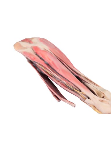
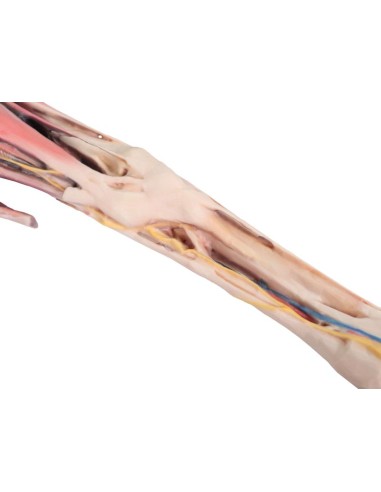
Realistic and Detailed Anatomy
- Accurate Representation: The model precisely reproduces the muscles, tendons, ligaments, vessels, and nerves of the horse’s forelimb, highlighting anatomical relationships from the distal region up to the knee joint.
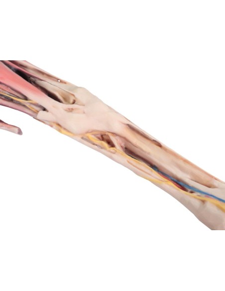
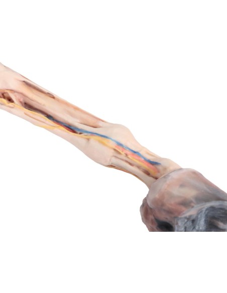
- 1:3 Scale: The 1:3 scale representation allows for magnified and detailed study, making even the smallest and most complex structures visible.
High-Quality Materials and Precision Finishing
- Selected Materials: Constructed from high-quality materials, the model ensures strength and durability, making it ideal for intensive use in educational and research settings.
- Attention to Detail: Each anatomical component is crafted with care, ensuring a realistic and precise representation, essential for effective learning.
Educational and Research Applications
- Advanced Educational Tool: Perfect for veterinary anatomy courses, the model allows students to gain practical and in-depth knowledge without the need to use live subjects.
- Research Support: Also useful for research projects, this tool facilitates comparative studies and detailed analysis of the anatomical structures of the horse’s forelimb.
Benefits of Use
Practical and Safe Learning
By using the Horse Forequarter Model (1:3 Scale), students and professionals can practice in a controlled environment, improving their understanding of complex anatomical structures and promoting an ethical approach to training without the use of live subjects.
Investment for Institutions and Research Centers
This model represents a strategic investment for veterinary schools, universities, and research centers, helping to elevate the level of education and clinical practice thanks to its high quality and advanced features.
Technical Specifications
For further information regarding dimensions, materials used, and instructions for use, we recommend consulting the complete technical data sheet or contacting our customer service for personalized assistance.
Buy Now
Don’t miss the opportunity to enrich your education or that of your students with the Horse Forelimb
Model – Muscles, Tendons, Ligaments, Vessels, and Distal Nerves up to the Knee Joint (1:3 Scale)
.
Add to cart
and discover how this tool can revolutionize the approach to studying veterinary anatomy.
What advantages does the Monash University anatomical dissection collection offer compared to plastic models or plastinated specimens?
- Each body replica has been carefully crafted from selected patient radiographic data or human cadaver specimens selected by a highly qualified team of anatomists at the Monash University Centre for Human Anatomy Education to illustrate a range of clinically important anatomical areas with a quality and fidelity that cannot be achieved with conventional anatomical models – this is real anatomy, not stylized.
- Each body replica has been rigorously checked by a team of highly qualified anatomists at the Center for Human Anatomy Education, Monash University, to ensure the anatomical accuracy of the final product.
- The body replicas are not real human tissue and are therefore not subject to any restrictions on transport, import, or use in educational institutions that do not hold an anatomy license. The Monash 3D Anatomy dissection series avoids these and other ethical issues that arise when dealing with plastinated human remains.
No reviews
Tap to zoom