Your cart
There are no more items in your cart


3B Scientific K26 Anatomical Model of Gallstones
3b scientific
1000314
Out-of-Stock
€76.50
Tax included
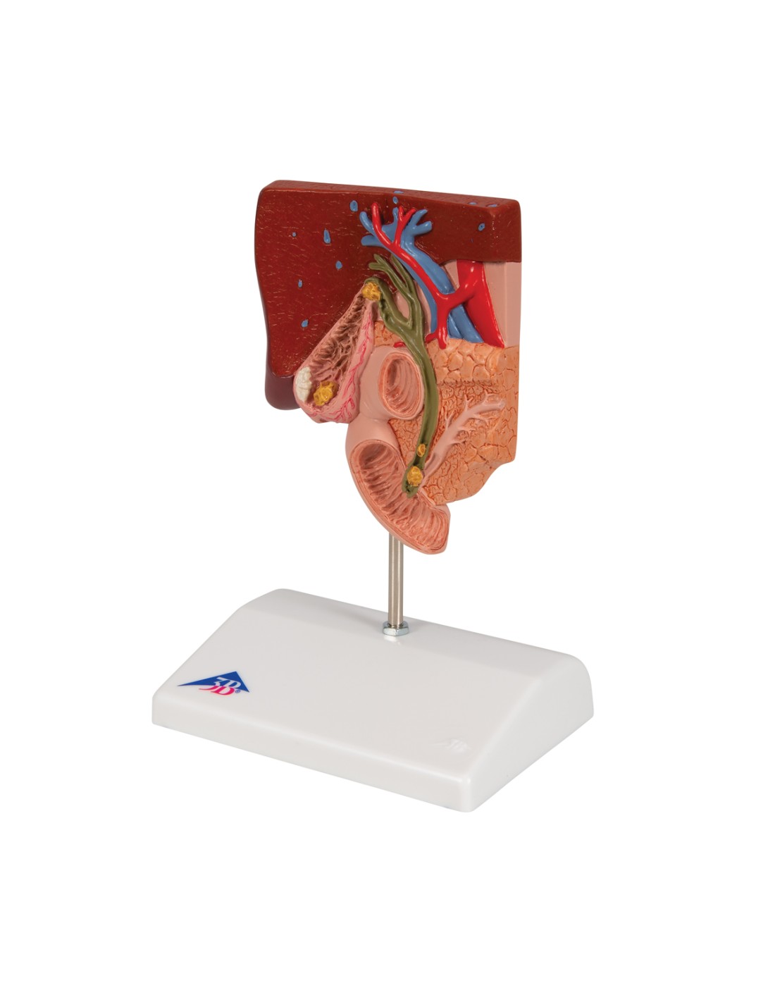
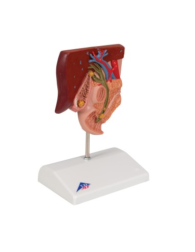
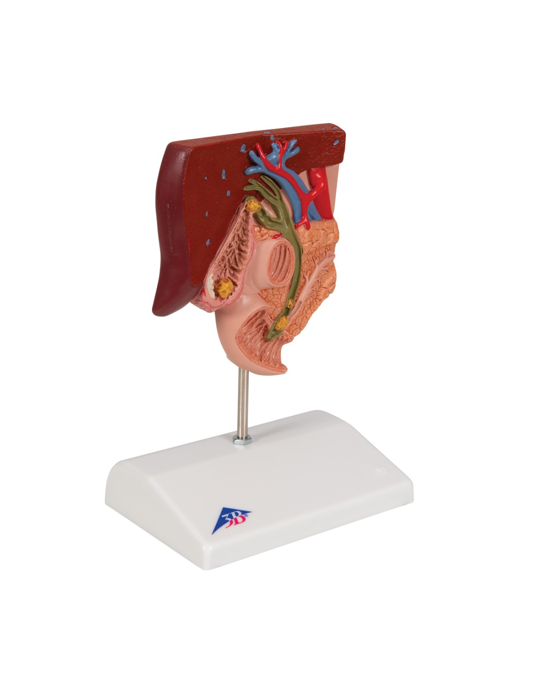
3B Scientific K26 Anatomical Model of Gallstones (1:2 scale). Demonstrates acute and chronic infections, with gallstones in four typical positions. Comes with a base featuring Smart Anatomy.
3B Scientific K26 Anatomical Model of Gallstones
Product Description
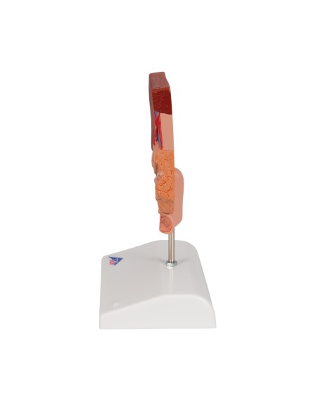
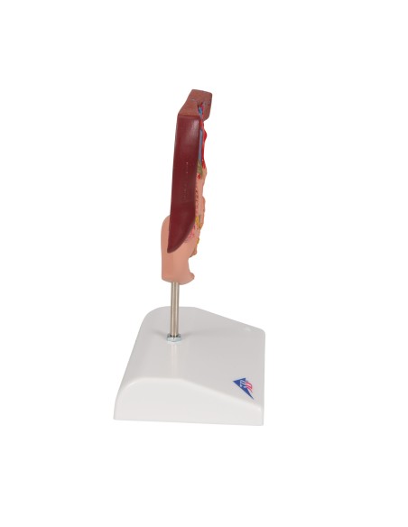
The 3B Scientific K26 anatomical model, scaled down to half the size of a real gallbladder, is designed to clearly illustrate the anatomy of the biliary system and the changes caused by the presence of gallstones.
Visible on the gallbladder wall are:
-
an acute infection (cholecystitis)
-
tissue changes due to a chronic infection
Gallstones are depicted in their typical sites of formation:
-
the base of the gallbladder
-
the area of the spiral valve
-
common bile duct
-
the papilla opening into the small intestine
The model is mounted on a base for practical and durable display, suitable for educational and clinical use.
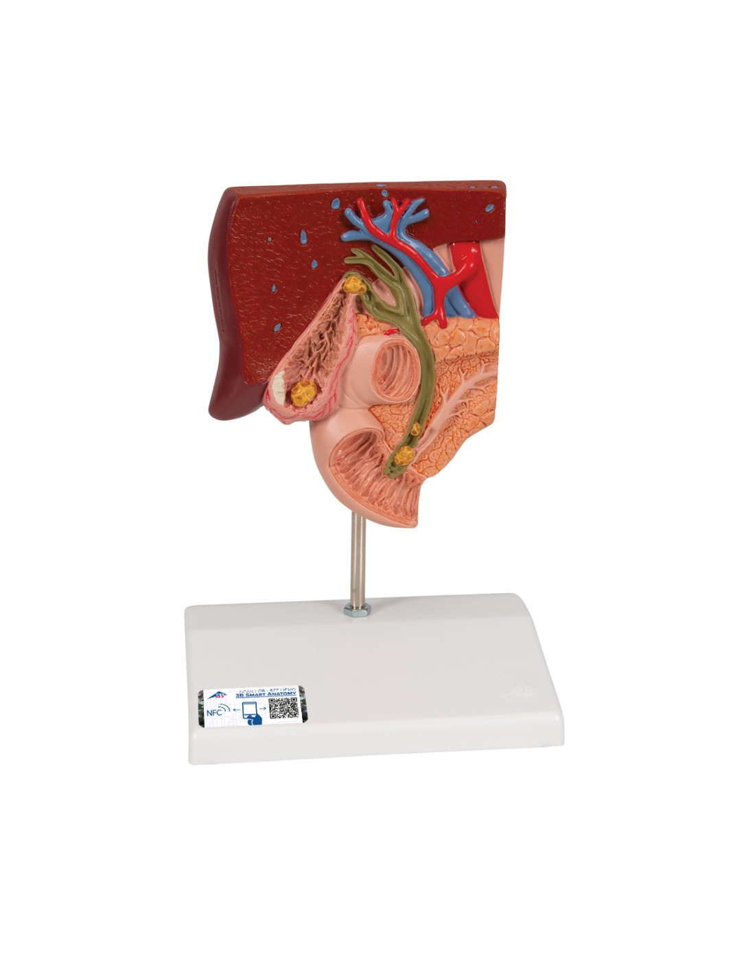
3B Smart Anatomy Digital Features
Every original 3B Scientific® anatomical model includes access to the interactive digital twin, usable on smartphones, tablets, and PCs, with advanced features:
-
3D visualization with zoom and free rotation
-
Augmented reality (AR) to place the model in real space
-
Anatomy quizzes with immediate results and final assessment
-
Customizable drawing and note-taking features
-
Online and offline access
-
Available in 11 languages
Technical specifications
-
Anatomical model of the biliary system (1:2 scale)
-
Demonstrates acute and chronic gallbladder infection
-
Gallstones depicted in 4 typical locations
-
Mounted on a base
-
Includes access to 3B Smart Anatomy
Educational and clinical applications
This model is an effective tool for explaining biliary disorders to students and patients. It is particularly useful in clinical settings to illustrate the causes and consequences of gallstones, as well as to demonstrate the effects of acute and chronic infections on the gallbladder.
- Height
- 15
- Width
- 26
- Depth
- 18,5
- Weight
- 1
No reviews
Tap to zoom