Your cart
There are no more items in your cart


3B Scientific K18 Anatomical Model of Esophageal Diseases
3b scientific
1000305
Out-of-Stock
€70.00
Tax included
3B Scientific K18 Anatomical Model of a Pathological Esophagus. Demonstrates reflux, ulcers, Barrett’s esophagus, carcinoma, varices, and hiatal hernia. Mounted on a base with 3B Smart Anatomy access.
Product Description
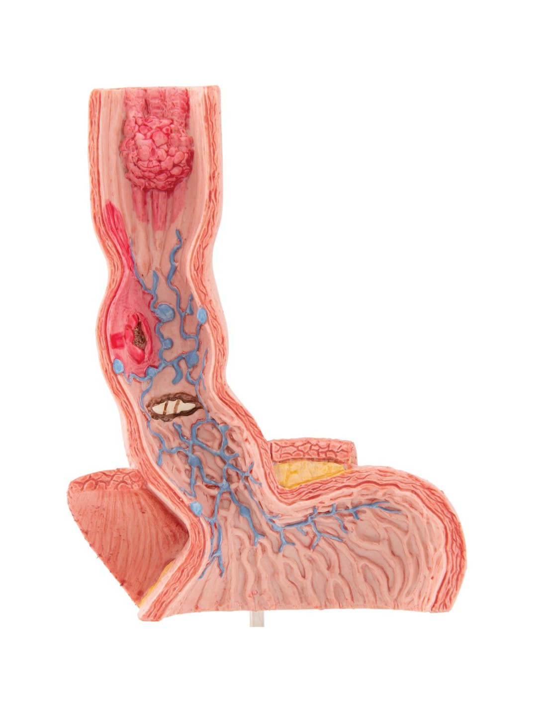
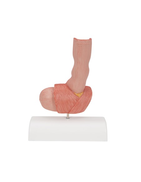
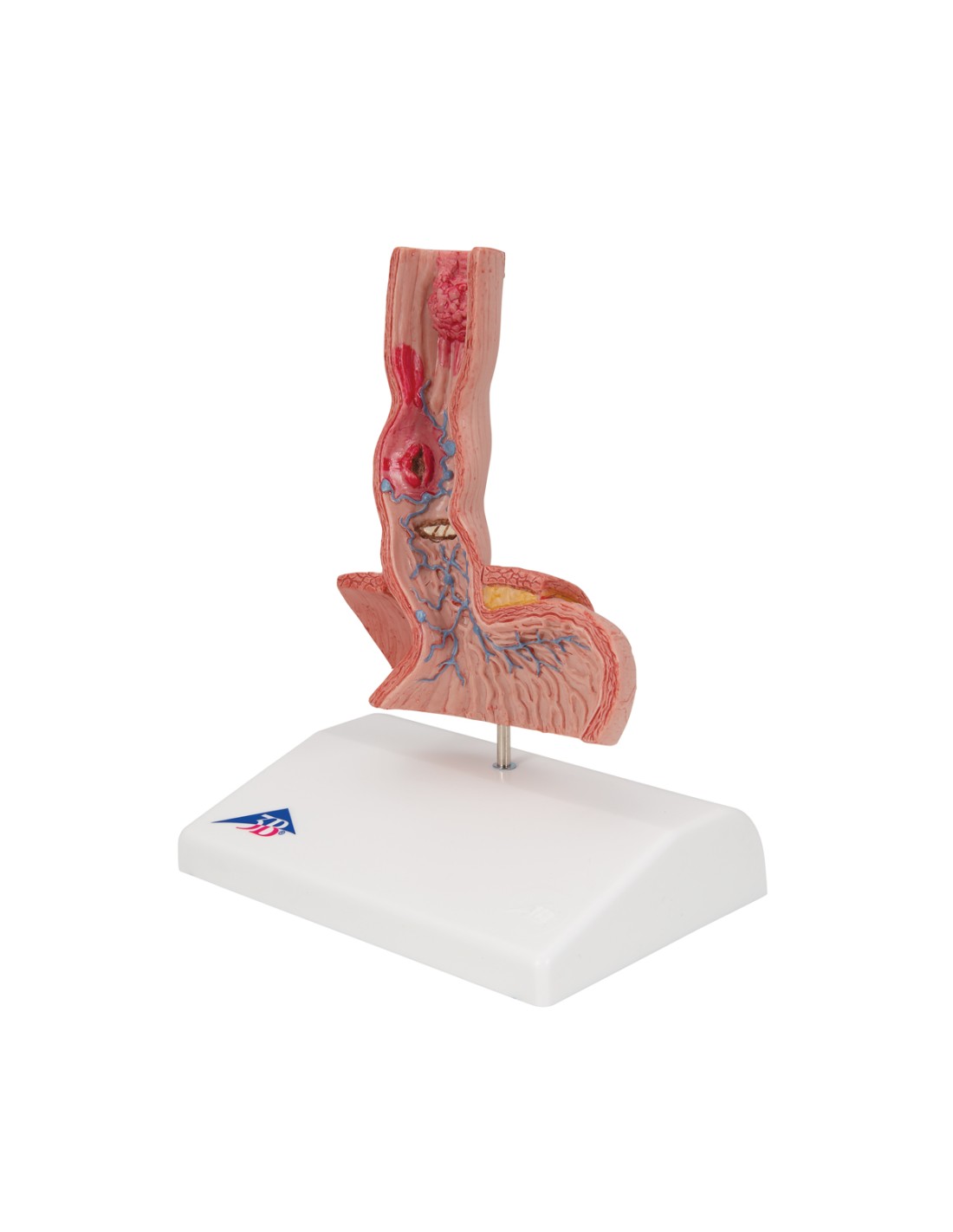
The 3B Scientific K18 anatomical model is designed to clearly and realistically illustrate the main pathologies of the esophagus, making it particularly useful in both educational and clinical settings.
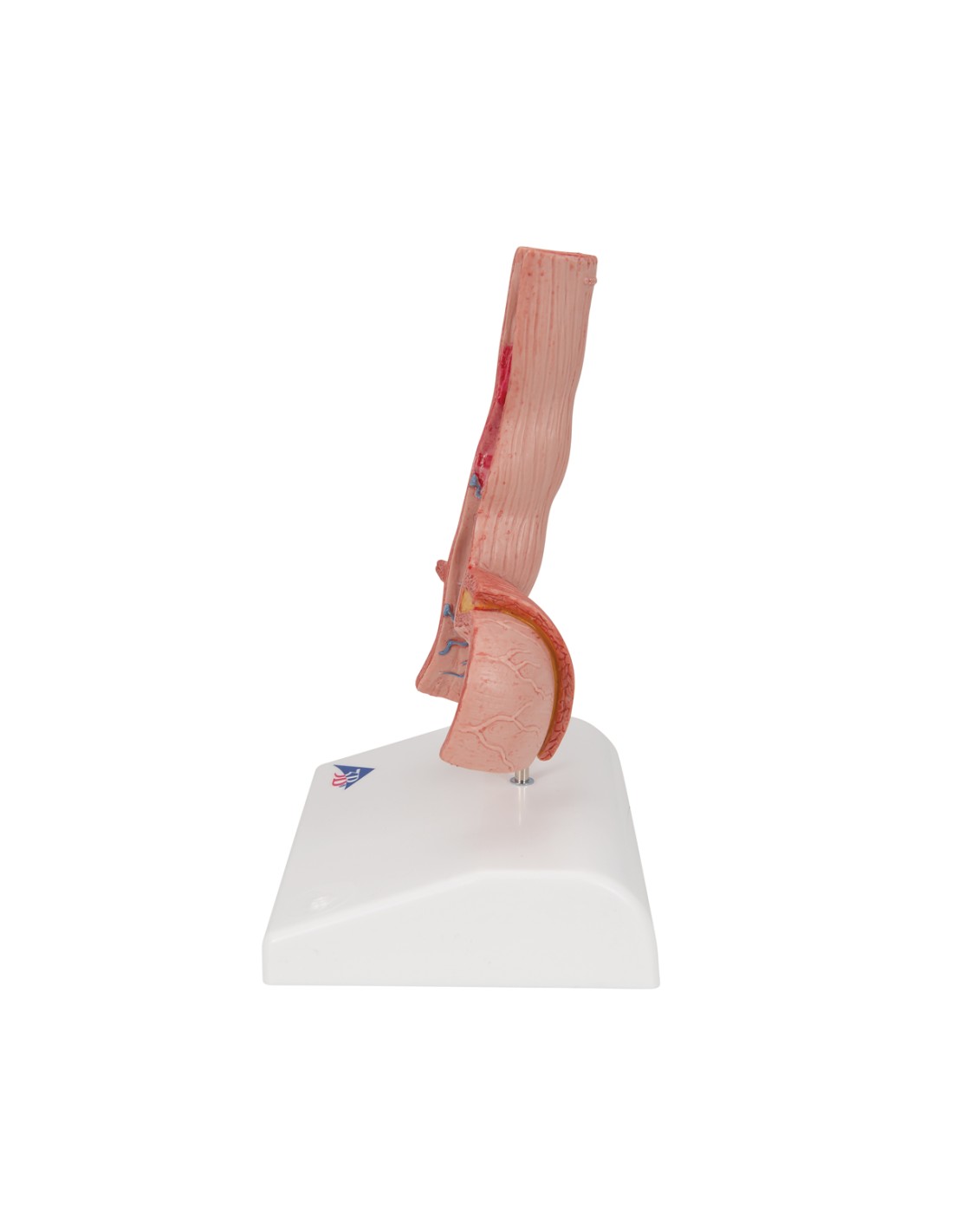
It is a life-size frontal section extending from the lower portion of the esophagus to the upper portion of the stomach, allowing for the observation of the most common pathological changes.
The diseases depicted are:
-
Reflux esophagitis
-
Esophageal ulcer
-
Barrett’s esophagus
-
Esophageal carcinoma
-
Esophageal varices
-
Hiatal hernia
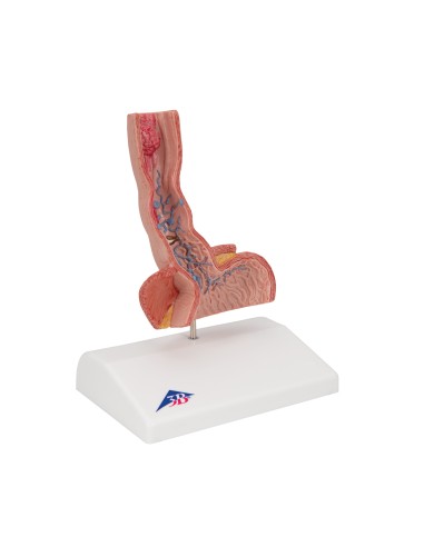
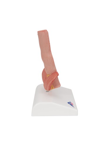
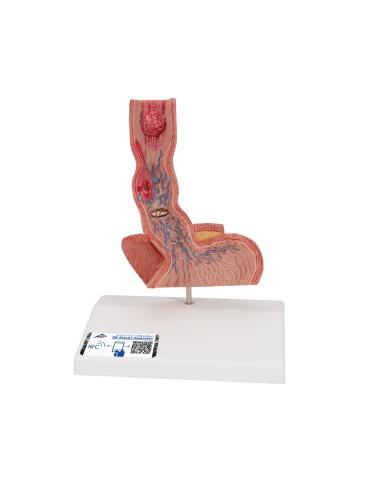
The model is mounted on a base for practical demonstration during classes, lectures, or patient consultations.
3B Smart Anatomy Digital Features
Every original 3B Scientific® anatomical model includes access to its interactive digital twin, usable on smartphones, tablets, and PCs, with the following features:
-
3D visualization with rotation, zoom, and interactive areas
-
Augmented reality (AR) activation
-
Anatomy quizzes with immediate scoring
-
Customizable drawing and note-taking features
-
Online and offline access to content
-
Available in 11 languages
Technical Specifications
-
Life-size frontal section model of the esophagus and upper stomach
-
Depicts: reflux esophagitis, ulcer, Barrett’s ulcer, esophageal carcinoma, esophageal varices, hiatal hernia
-
Mounted on a base
-
Access to 3B Smart Anatomy digital content included
No reviews
Tap to zoom