
Your cart
There are no more items in your cart

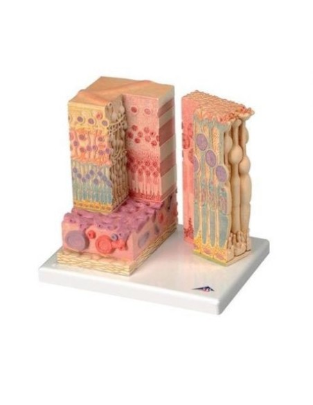
3B Scientific, 3B MICROanatomy F16 anatomical model of eye tissues.
3b scientific
3401
Last items in stock
€249.90
Tax included
Based on
3B scientific anatomical models, anatomical model of eye tissues F16
Anatomical model of eye, magnified 3 times, decomposable into 6 parts,on base.
This extraordinary anatomical model illustrates the structure of the fine tissues of the retina with sclera and choroid.
The left section of the model, a stepped block, shows the entire structure of the retina from a microscopic point of view, with the afferent vessel layer and parts of the sclera.
In contrast, the right section of the model is an enlarged detail, illustrating the fine structure of the photoreceptors and pigment epithelium cells.
Left section magnified 850 times - right section magnified 3800 times.
Based on.
3B Scientific F16 anatomical eye model is made of polyvinyl chloride (PVC), which is very hard, lightweight and corrosion resistant. PVC resists reactions with acids, alcohol gasoline, and hydrocarbons.
Why buy it
3B Scientific Anatomical Models are undoubtedly the best on the market, ideal for teaching, providing clarification to patients, and scientific medical training.
Many physicians and professionals purchase the anatomical teaching models to highlight key points on topics such as the skeleton, human musculature, joints and related diseases (rheumatism, arthritis, bursitis, synovitis, bursitis, tendonitis, cervical, ischialgia).
They can also be used as furnishing accessories to personalize one's medical office.
- Height
- 25
- Width
- 23
- Depth
- 18,5
- Weight
- 1.2
No reviews
Tap to zoom
